To Be Hale and Hearty, Brain Microglia Need a Healthy Gut
Quick Links
The human intestine contains a rich mosaic of microbial cells—around 1,000 different species—that help digest food, block pathogens, and maintain general good health. A paper in the June 1 Nature Neuroscience assigns the gut microbiome yet another role. Researchers led by Marco Prinz, University of Freiburg, Germany, found that these microorganisms help mature and bolster the brain’s resident macrophages, known as microglia. The connection appears to hinge on short-chain fatty acids produced by the bacteria as they digest food. While there are no obvious implications yet for neurodegeneration, microglia are increasingly recognized as important players in diseases such as Alzheimer’s and Parkinson’s. Grasping the cells’ basic biology could offer clues to these disorders.
“Understanding more about what regulates the maturation, growth, and responses of microglia within the CNS is a critically important area,” said Terrence Town, University of Southern California, Los Angeles, who was not involved in the study. “This study illustrates the dynamic and complex interplay between gut flora and microglia—not an obvious connection.”
Scientists already knew that gut flora influenced a variety of peripheral immune cells. Mice raised without microbiota—known as germ-free—have immature and underperforming immune systems (for a review, see Round and Mazmanian, 2009). However, no one had explored whether microorganisms in the intestine help mature and maintain the brain’s immune cells, which are locked securely away behind the blood-brain barrier.
To find out, co-first authors Daniel Erny and Anna Lena Hrabě de Angelis compared microglia in germ-free mice with those raised in mice with normal intestinal bacteria. All the mice were six to 10 weeks old. According to deep sequencing, microglial mRNA profiles differed dramatically between the two sets of mice. In germ-free animals, 198 genes were downregulated, including those for cell activation, pathogen recognition, and signaling. Moreover, 173 genes that normally taper off in mature microglia were upregulated, including those important for transcriptional regulation, survival, and proliferation. Surface molecules typical of immature microglia also dotted their cell membranes. Together, the results suggest microglia in germ-free mice were stalled in a juvenile state.
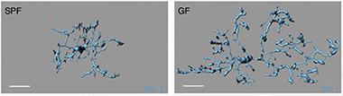
Malformed Microglia:
Microglia from a mouse with no germs in its gut (right) dwarf those from mice with a healthy microbiome (left). [Image courtesy of Erny et al., Nature Neuroscience.]
Histopathological and morphological findings corroborated that result. Brains of germ-free mice hosted much higher numbers of microglia, with longer processes, more segments, and greater numbers of branching and terminal points (see image). They bumped up against one another more often, whereas normal microglia held to stricter boundaries.
It wasn’t just the cells’ maturation and appearance that took a hit. Their reaction to infection also faltered. If the researchers simulated a bacterial infection using lipopolysaccharide, the malformed microglia mounted a weaker response than those in control animals. The same held true when the researchers injected lymphocytic choriomeningitis virus into a mouse’s brain. Germ-free microglia multiplied more slowly and responded less robustly than in healthy mice.
Interestingly, microglial maintenance seems to depend on continuous input from gut microorganisms. If the scientists wiped out intestinal microorganisms of healthy mice with antibiotics, microglia assumed the shape and immaturity of microglia in germ-free animals. Microglia also seem to require a full range of microorganisms. Adding a few select strains to the intestines of germ-free mice did nothing to restore microglia to health, but colonizing mice with a complex array of gut flora normalized their numbers and largely restored their shape.
What connects gut microorganisms to the far-off brain? The researchers suspected short-chain fatty acids. Gut bacteria produce SCFAs as byproducts of fermentation, and they were recently reported as stabilizing immune cells in the colon (Smith et al., 2013). Sure enough, simply adding SCFAs to the drinking water of germ-free mice for four weeks returned microglial shape, maturity, and number almost back to normal.
In mice that lacked the GPR43 receptor known to recognize SCFAs, microglia retained their malformed state. However, the researchers could not find this receptor anywhere in the brains of these mice. It could be that SFCAs act in the periphery to influence the brain, or that a different receptor recognizes them in the central nervous system, said Town. He noted that SFCAs are known to cross the blood-brain barrier.
All in all, the data suggest that an important connection persists between gut flora and the brain’s immune system throughout life, wrote the authors. Town proposed that the researchers next test whether SFCAs restore microglial function.
Does this study mean anything for Alzheimer’s? Patients with the disease are known to become malnourished (Magri et al., 2003). Town speculated that they may have an impoverished microbiome, which could exacerbate disease.—Gwyneth Dickey Zakaib
References
Paper Citations
- Round JL, Mazmanian SK. The gut microbiota shapes intestinal immune responses during health and disease. Nat Rev Immunol. 2009 May;9(5):313-23. PubMed.
- Smith PM, Howitt MR, Panikov N, Michaud M, Gallini CA, Bohlooly-Y M, Glickman JN, Garrett WS. The microbial metabolites, short-chain fatty acids, regulate colonic Treg cell homeostasis. Science. 2013 Aug 2;341(6145):569-73. Epub 2013 Jul 4 PubMed.
- Magri F, Borza A, del Vecchio S, Chytiris S, Cuzzoni G, Busconi L, Rebesco A, Ferrari E. Nutritional assessment of demented patients: a descriptive study. Aging Clin Exp Res. 2003 Apr;15(2):148-53. PubMed.
Further Reading
Papers
- Shoemark DK, Allen SJ. The microbiome and disease: reviewing the links between the oral microbiome, aging, and Alzheimer's disease. J Alzheimers Dis. 2015;43(3):725-38. PubMed.
- Yadav SK, Mindur JE, Ito K, Dhib-Jalbut S. Advances in the immunopathogenesis of multiple sclerosis. Curr Opin Neurol. 2015 Jun;28(3):206-19. PubMed.
- Astafurov K, Elhawy E, Ren L, Dong CQ, Igboin C, Hyman L, Griffen A, Mittag T, Danias J. Oral microbiome link to neurodegeneration in glaucoma. PLoS One. 2014;9(9):e104416. Epub 2014 Sep 2 PubMed.
- Bereswill S, Kühl AA, Alutis M, Fischer A, Möhle L, Struck D, Liesenfeld O, Göbel UB, Dunay IR, Heimesaat MM. The impact of Toll-like-receptor-9 on intestinal microbiota composition and extra-intestinal sequelae in experimental Toxoplasma gondii induced ileitis. Gut Pathog. 2014;6:19. Epub 2014 Jun 6 PubMed.
- Magnusson KR, Hauck L, Jeffrey BM, Elias V, Humphrey A, Nath R, Perrone A, Bermudez LE. Relationships between diet-related changes in the gut microbiome and cognitive flexibility. Neuroscience. 2015 May 14;300:128-140. PubMed.
- Mosher KI, Wyss-Coray T. Go with your gut: microbiota meet microglia. Nat Neurosci. 2015 Jun 25;18(7):930-1. PubMed.
Primary Papers
- Erny D, Hrabě de Angelis AL, Jaitin D, Wieghofer P, Staszewski O, David E, Keren-Shaul H, Mahlakoiv T, Jakobshagen K, Buch T, Schwierzeck V, Utermöhlen O, Chun E, Garrett WS, McCoy KD, Diefenbach A, Staeheli P, Stecher B, Amit I, Prinz M. Host microbiota constantly control maturation and function of microglia in the CNS. Nat Neurosci. 2015 Jun 1; PubMed.
Annotate
To make an annotation you must Login or Register.
Comments
University of New Mexico
This is a fascinating study, which conclusively links the gut microbiome to microglial homeostasis. The immune challenges and the models used are quite appropriate. With this and many recent studies (for example: Louveau et al., 2015), the concept of the brain being “immune-privileged” is fading away as many of the regulators of peripheral immune function (including the gut microbiome) are constantly influencing microglial activation and function within the CNS.
While this study opens up a whole new arena of research on the influence of the microbiome on microglial function, it also opened a lot of questions, including: What is the indirect effect of altered microglial function on neuronal homeostasis (e.g., microglia-mediated synaptic pruning and plasticity) and function? What is the role of SCFAs derived from a commensal “healthy” microbiome versus the ones derived following bacterial infection?
References:
Louveau A, Smirnov I, Keyes TJ, Eccles JD, Rouhani SJ, Peske JD, Derecki NC, Castle D, Mandell JW, Lee KS, Harris TH, Kipnis J. Structural and functional features of central nervous system lymphatic vessels. Nature. 2015 Jul 16;523(7560):337-41. Epub 2015 Jun 1 PubMed.
This is an important study that indicates how important the gut microcbiome is to brain health via microglial activation. Further investigation into SCFA food sources and their impact on disease processes/immunology should prove very interesting to budding connections in the field of gastroenteroneuroimmunology.
Make a Comment
To make a comment you must login or register.