Cortical Hubs to Rich Clubs—Linking Brain Connectivity to Function
( 3 Articles Available, 0 Articles Pending )
If you find it hard to keep up with Facebook, LinkedIn, and other social networks, spare a thought for the brain. With 100 to 500 trillion synaptic connections, the human brain dwarfs all of them. How do those connections work to formulate thought, recall memories, or perform any basic brain function? An expanding repertoire of neuroimaging and molecular techniques is empowering scientists to delve into this mass of activity to unpack elaborate processes.
As reported in a number of recent papers, researchers are finding an uncanny degree of interlinkage between a set of cortical regions; gaining insight into how network patterns change with time; correlating memory function with structural connectivity and electrical oscillations; and identifying neural networks that underlie specific branches of memory.
Cortical Hubs Form "Rich Club" in Human Brain
With 100 billion neurons, each signaling to roughly 7,000 others, the human brain bustles with mind-boggling activity. Yet within the hum of these sprawling networks, scientists are teasing out structural and electrophysiological underpinnings of an exquisitely complex process—memory. Using magnetic resonance imaging (MRI) and electroencephalography (EEG), researchers are finding what they call a “rich club”—a set of unusually interconnected brain regions—and learning how network patterns change with time (Part 1). Other brain imaging research ties memory function with structural connectivity and electrical oscillations in defined cortical regions (see Part 2). And additional studies—one using transgenic mice—home in on specific neural networks and show how their dysfunction weakens memory (see Part 3).
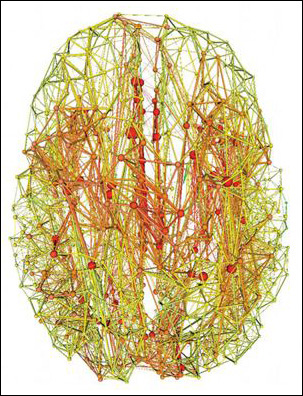
Just as certain individuals know lots of people and keep everyone up to date with the latest news, a small number of cortical “hubs” hook up with many other brain areas to coordinate the flow of information among them. Researchers have used MRI to correlate neural activity across these hubs (ARF related news story on Buckner et al., 2009) and trace physical connections among them (Hagmann et al., 2008), building a map of their connectivity. The hubs appear in several cortical subregions, including the posterior cingulate, lateral temporal, lateral parietal, and medial/lateral prefrontal areas (regions among the earliest affected in Alzheimer’s disease). In the November 2 Journal of Neuroscience, Martijn van den Heuvel of University Medical Center Utrecht, The Netherlands, and Olaf Sporns of Indiana University, Bloomington, took things a step further. Analyzing diffusion tensor imaging data from 21 young adults, the researchers discovered that brain hubs not only have more total connections, but “they tend to have more connections with their hub friends…in that sense, forming a collective, or so-called ‘rich club,’ in the brain,” van den Heuvel told ARF (see image below). He estimated that the hubs have 1.5 to two times more connections among each other than would be expected based on pure chance. “Having double the number of connections you should have is quite extensive,” van den Heuvel said.
Magnetic resonance imaging data reveals areas of the human brain with the fewest (green) and greatest (red) number of connections. Well-connected “hubs” have a disproportionately high number of connections with each other. Image credit: Van den Heuvel et al. Journal of Neuroscience 2011
That got the scientists thinking about the potential impact of these dense hubs on disease. If likened to electric power grids, the rich club might “give a level of robustness to the network,” van den Heuvel said. “If one station fails, the others can easily take over because they are interconnected.” However, having a high concentration of connections carries some risk, too. As with the electric grid, if a major hub “goes down, it can take down others as well.” In a hypothetical model of the impact of targeted and random damage to neuronal networks, the authors determined that specific attacks on rich-club connections slowed information flow about three times more than harm to random parts of the network.
The findings may be relevant to Alzheimer’s disease. It is known that cortical hubs are hot spots of amyloid-β, and the rich club encompasses areas of the hippocampus, which is critical for memory and highly susceptible to AD pathology. “Alzheimer’s disease may, therefore, disproportionately affect the ability of other regions of the brain to interact with the hippocampus through its rich-club connections,” noted Trey Hedden of Massachusetts General Hospital, Charlestown, in an e-mail to ARF. The authors plan to apply their brain imaging protocol to AD and schizophrenia patients—“to see if their ‘rich club’ is less connected in memory areas,” van den Heuvel said.
The rich-club arrangement—where hubs associate more with each other than with less connected centers in a network—had already been described in other walks of life, such as the Internet. When the cortical hubs were mapped several years ago, Sporns and colleagues wondered whether they formed a rich club in the brain as well. The current study fits into a larger line of research aimed at producing a comprehensive map of all possible neuronal connections in the human brain (see Sporns, 2011 review), van den Heuvel noted. The Human Connectome Project is creating an interface to navigate such data.
Reporting in the same issue of JNS, researchers led by Mark Kramer of Boston University and Sydney Cash of MGH, Boston, also took a hard look at the brain’s structural layout—but with the added dimension of time. From 24-hour intracranial EEG recordings in six epileptic patients already undergoing invasive monitoring as part of their treatment, the scientists constructed functional networks and analyzed how they evolve over time. “What we found was moment-to-moment variability, yet a few functional network patterns that reappeared again and again,” Kramer noted in an e-mail to ARF. Granted, the intracranial EEG recordings only cover a limited part of the brain, and the spatial resolution of this technique is about 10 times worse than fMRI. On the flip side, fMRI has poorer temporal resolution (seconds rather than milliseconds in EEG), and practical considerations including scanner cost and access prohibit long-term fMRI recording. In the current study, similar network characteristics came up for all six patients despite differences in age, sex, and disease etiology, lending credence to the analysis. Furthermore, the findings seemed consistent with scalp EEG recordings in healthy people and in task-related EEG. The data suggest that just 100 seconds of continuous recording can provide “an indication of the brain’s functional connectivity network with high fidelity,” the authors write.
If a “metastable” pattern of brain interactions continually appear, Kramer speculates, “we can attempt to deduce these potential patterns of interaction from a few minutes of brain voltage data, and determine how these patterns alter during the course of aging or neurodegeneration.”—Esther Landhuis
Just about everyone knows a person who seems to know an unusually large number of people, has the latest information on how they are doing, and can quickly put them in touch with a potential friend or colleague. In the study of social network theory, these people are known as "hubs" or "connectors." Similarly, the human brain appears to be organized so that a small number of key brain regions have many connections to other regions of the brain and likely act as hubs to facilitate the transfer of information. This pattern of connectivity can be measured by examining the functional connections among brain regions (measuring how their activity is spontaneously correlated; see Buckner et al., 2009), or by examining the structural connections via which neural signals are transmitted. The paper by van den Heuvel and Sporns uses a technique to measure the white matter fiber tracts that make up the structural connectivity of the brain.
They find that a small set of brain regions have both a high degree of interconnectedness with other regions of the brain and a high degree of crosstalk with one another. This they term a so-called "rich club" (regions rich in connections that meet together in a semi-private club). Not surprisingly, these rich-club regions happen to be key structures in the relay of information (e.g., the thalamus has long been known to relay sensory information to cortical regions) or are positioned at junctures of major white matter fiber tracts (e.g., the precuneus is positioned at a juncture of the corpus callosum, which connects the brain's hemispheres, and the superior longitudinal fasciculus, which connects the frontal lobe with the parietal and occipital lobes).
They model the effects of selectively damaging the interconnections between these rich-club regions and find that such damage is particularly devastating to the shortest-path connections between two regions. In other words, when rich-club regions are severed from one another, information can only flow between regions along much less efficient routes. It is important to point out that this is a hypothetical model only, as such severe trauma to the densest white matter tracts is rare and usually only seen in cases of last-resort surgery. The brain is also a highly adaptive organ, and can partially "rewire" itself in response to milder trauma.
Of interest to the study of Alzheimer's disease, one of the regions that is a member of the rich-club, the hippocampus, is both central to memory formation and highly susceptible to the neurofibrillary tangles found in AD. AD may therefore disproportionately affect the ability of other regions of the brain to interact with the hippocampus through its rich-club connections. Other work has shown that there is a high degree of overlap in the accumulation of amyloid plaques and areas of the neocortex with a high degree of connectivity (including many of the same areas identified here as the rich-club), so AD may have a second route of impact by destroying the ability of these neocortical regions to interact with one another and with the hippocampus.
Wired for Memory? Electrical and Structural Connectivity Linked
Beyond flash cards and mnemonics, hard-wired differences in brain structure and neural firing rhythms may give some people an edge in their ability to remember things. That’s the gist of a study, published in the November 3 Current Biology online, which used a combination of brain imaging techniques to correlate memory function with structural connectivity between the prefrontal cortex (PFC) and the hippocampus. In another study—described in the November 2 Journal of Neuroscience—researchers mapped electrical oscillations underlying memory formation to the PFC as well. The findings strengthen the idea that oscillatory signals detected during memory formation may predict the success of later recall.
Traditionally, scientists have pegged the prefrontal cortex as the brain’s working memory center, holding information needed for complex reasoning and learning, while they attribute long-term memory to the hippocampus. As it turns out, connections between these two brain regions drive both types of memory, yet studying the structures in concert in living people is challenging.
In the Current Biology paper, Michael Cohen of the University of Amsterdam, The Netherlands, designed a dual-pronged approach to study how the hippocampus and prefrontal cortex contribute to memory. In one set of experiments, Cohen used a type of MRI called diffusion-weighted imaging to trace white matter connections between the prefrontal cortex and hippocampus in 20 healthy, young adults. “This has nothing to do with function; it’s all about structure. It’s measuring a quality of their brain that is physically there all the time,” Cohen told ARF. Then, in a separate analysis done on the same people a week or two later, he used electroencephalography (EEG) to record prefrontal cortex activity during a difficult memory task. Study participants saw a series of line drawings flash momentarily onto a computer screen, then had to remember whether the drawing was rotated slightly clockwise or counterclockwise when it reappeared five seconds later. Long-term memory was tested about an hour later, when the participants viewed some of the original pictures along with deceptively similar “lures," and indicated whether they thought they had previously seen the drawing.
“People with stronger connectivity (measured by MRI) between the hippocampus and prefrontal cortex were better able to encode the pictures and remember them,” Cohen said. “These are fundamental, hard-wired, physical features of your brain.”
Though the study’s main focus was linking memory performance to structural connectivity, Cohen noted, the subjects with stronger hippocampus-PFC connections also had slower task-related brain oscillations, as measured by the EEG. Oscillations are rhythmic fluctuations in brain activity that reflect the excitability of large cell populations. Measuring this electrical activity allows scientists to gauge how well the brain stores and transmits information from one region to another. The present data confirm earlier theoretical studies suggesting that slow oscillations play a key role in memory maintenance, Cohen said.
The present study also represents a methodological advance. By linking a fairly old method (EEG) with a more recent technique (diffusion-tensor MRI), the multimodal approach links brain structure to function and “helps overcome one of the main limitations of EEG: the inability to measure deep brain structures,” Cohen wrote. Ongoing work in his lab is addressing whether age or other factors such as learning might influence the strength of hippocampal-PFC connectivity.
Rather than correlate brain function with underlying structure, the Journal of Neuroscience study linked memory function with EEG oscillations at signature frequencies, namely β (13-30 Hz) and ϑ (4-8 Hz). The analysis began as an attempt to replicate counterintuitive results of a previous study, said first author Simon Hanslmayr of the University of Konstanz, Germany. In that study (Hanslmayr et al., 2009), he and senior author Karl-Heinz Bäuml linked semantic memory with a decrease in β oscillations. This was unexpected because less β power means less synchronous neuronal firing, and “we had very little idea how that could serve memory encoding,” Hanslmayr told ARF. It made more sense that stronger synaptic connections, measured as greater synchrony, or increased β power, drove memory, he noted.
In the current paper, Hanslmayr and colleagues not only confirmed their 2009 findings, but also mapped the decrease in β synchrony to a specific brain area—the left inferior frontal gyrus. “It’s not just background noise,” Hanslmayr said. His team found this by doing simultaneous EEG and functional MRI (fMRI) on 24 healthy volunteers (mean age 23 years) while they memorized word lists. For each word, the researchers noted whether the volunteers successfully recalled it, then looked at their blood oxygen level-dependent (BOLD) fMRI changes to see which part of the brain was active during encoding of words that were later recalled successfully. They found that BOLD signal was up, and β power down, in the left inferior frontal gyrus, but only for words the volunteer remembered. The scientists saw no such correlation for items that were not recalled.
Alongside the decline in β signal, the scientists also found that good memory correlated with increased ϑ oscillations in medial temporal lobe regions, consistent with prior studies (Rutishauser et al., 2010; see also review by Axmacher et al., 2006).
The paper shows that BOLD signal and electrophysiology are correlated only when subjects are attentively processing the items, presumably leading to good memory later, Hanslmayr said. And, reassuringly, Cohen said the “regions showing the oscillatory signatures are in the same part of the PFC that had strong connectivity with the hippocampus and predicted subjects’ memory [in our study].” “I think a very important message of our paper is that researchers should not neglect the middle-range frequencies (like β and α oscillations) and should really look at power decreases,” said Hanslmayr. He added that the mid-range frequencies are important for long-term memory.
On the methodological side, Hanslmayr’s research suggests that β power measured by EEG may be able to serve as a proxy for memory encoding, without need for fMRI—a scanning procedure that can be hard to perform on ailing elderly or those with dementia. Hanslmayr hopes his paper will inspire other scientists to look more closely at power decreases in the middle-range frequencies, e.g., β and α oscillations, and their role in long-term memory.—Esther Landhuis
Seeing With the Mind’s Eye—Not So Easy for Seniors
If asked to describe what you ate for dinner last night, chances are you would picture the meal in your mind before responding. Older adults have trouble with these sorts of mental imagery tasks. A functional brain imaging study in the November 2 Journal of Neuroscience offers insight into this deficit—the aging mind’s vision doesn’t shut down; it gets blurred, the study suggests. Moreover, this age-related imagery decline seems to correlate with poor visual memory, suggesting the brain really needs to “see” what it is trying to recall. In another recent advance reported in the November 3 Science, researchers blocked synaptic transmission along certain circuits in transgenic mice, allowing the scientists to probe neural pathways underlying specific types of memory. The two studies are representative of the progress neuroscientists are making in understanding learning and memory.
When people look at something or picture it in their minds, neurons in the visual cortex start firing, but that’s not all. Mental imagery also activates prefrontal cortical areas that help the brain process information in a goal-oriented manner, by focusing on relevant stimuli and ignoring distractions in a process known as top-down modulation (Kosslyn et al., 1997; Ishai et al., 2000). This capability underlies spatial navigation and other visual memory functions that tend to worsen with age. In behavioral studies, seniors who performed more slowly and less accurately in memory tasks that required mental imagery reported having problems visualizing images in their minds (Briggs et al., 1999). “What was missing was quantitative neural data that spoke to this [subjective report],” said lead investigator Adam Gazzaley of the University of California, San Francisco.
As described in their paper, first author Jonathan Kalkstein and colleagues used functional magnetic resonance imaging (fMRI) to measure brain activity in 14 seniors (average age 69.8 years) and 14 young people (average age 26.6 years) as they fixated on moving images. While inside the MRI scanner, subjects saw a series of pictures—either celebrity faces or moving dots. In later “imagine” trials, the participants did not see the images but were instead prompted to visualize them.
During the “view” series, fMRI blood oxygen level-dependent (BOLD) signal went up in the fusiform gyrus—the facial recognition center—when faces were shown, and in the middle temporal gyrus when the images were moving dots. Activity in these visual areas was about the same in older and younger adults as they viewed actual images.
However, differences cropped up when participants were asked to picture those images in their minds. If prompted to imagine a celebrity’s face, young participants activated the brain’s face-selective area, but not the motion-selective area—and if asked to picture the moving dots, the latter area lit up, but not the former. The older participants, on the other hand, activated both areas of the visual cortex no matter what they were prompted to visualize. Like the young participants, “older adults were generating visual activity while they were imagining, but the big difference is they were not doing it selectively,” Gazzaley said. “This is evidence that older adults are not achieving as fine a resolution of the images they’re trying to create in their minds.”
Furthermore, the age-related imagery deficit seems to correlate with a drop in memory performance. Older participants were given the Continuous Visual Memory Test (CVMT). The lower-performing half had poorer motion imagery relative to young adults, whereas seniors with above-median CVMT scores did not have this visual imagery deficit.
Gazzaley said he hopes to design an adaptive training program to see if promoting mental imagery in seniors could improve visual memory.
Mouse Models—Dissecting Neural Pathways in Finer Detail
While fMRI offers scientists a powerful glimpse at the brain areas that mediate certain kinds of memory, figuring out precisely which neural networks are involved requires fine manipulations that are off limits in people—but could be done in mouse models. In an online Science paper published November 3, Susumu Tonegawa of the Picower Institute for Learning and Memory at MIT and colleagues generated a transgenic mouse to show that temporal association memory requires a specific branch of the hippocampal-entorhinal network.
First author Junghyup Suh and colleagues used a triple-transgenic strategy developed in the Tonegawa lab to block neurotransmitter release from a specific type of presynaptic cell in a temporally controlled manner. The “block” is carried out by tetanus toxin light chain (TeTxLC), which cleaves a protein (VAMP2) needed for fusion of synaptic vesicles with the presynaptic membrane. However, through transgenic crosses, expression of the toxin is tightly restricted to cells where two different promoters are active—in this case, one (Oxr1) that drives expression in entorhinal cortex layer III, and another (CamKII) that is active in excitatory neurons. The system has yet another level of control—doxycycline, which blocks toxin expression, overriding the activity of both promoters. In the present study, the researchers maintained the mice on a doxycycline diet until a month or two before doing behavioral tests. When the animals were switched to a doxycycline-free diet, tetanus toxin became expressed only at entorhinal cortex layer III inputs to the hippocampus, thereby achieving selective inhibition of synaptic transmission along this pathway.
Triple-transgenic mice with this selective block had no obvious molecular abnormalities in the entorhinal cortex, and looked normal in tests of anxiety, motor coordination, and pain sensitivity. However, the mice had problems with fear conditioning and temporal association memory, or the ability to link one event with another in time. Several years ago, Tonegawa’s group used this triple-transgenic approach to block synaptic transmission from the CA3 hippocampal area into the entorhinal cortex, and showed that this pathway is required for fast, one-trial contextual learning (Nakashiba et al., 2008). The team is trying to further delineate the neural circuits needed for spatial working memory and fear conditioning by using optogenetic methods to stimulate or inhibit specific inputs from entorhinal cortex to hippocampus in a much finer temporal window, Suh wrote in an e-mail to ARF.—Esther Landhuis
Comments
Massachusetts General Hospital
Just about everyone knows a person who seems to know an unusually large number of people, has the latest information on how they are doing, and can quickly put them in touch with a potential friend or colleague. In the study of social network theory, these people are known as "hubs" or "connectors." Similarly, the human brain appears to be organized so that a small number of key brain regions have many connections to other regions of the brain and likely act as hubs to facilitate the transfer of information. This pattern of connectivity can be measured by examining the functional connections among brain regions (measuring how their activity is spontaneously correlated; see Buckner et al., 2009), or by examining the structural connections via which neural signals are transmitted. The paper by van den Heuvel and Sporns uses a technique to measure the white matter fiber tracts that make up the structural connectivity of the brain.
They find that a small set of brain regions have both a high degree of interconnectedness with other regions of the brain and a high degree of crosstalk with one another. This they term a so-called "rich club" (regions rich in connections that meet together in a semi-private club). Not surprisingly, these rich-club regions happen to be key structures in the relay of information (e.g., the thalamus has long been known to relay sensory information to cortical regions) or are positioned at junctures of major white matter fiber tracts (e.g., the precuneus is positioned at a juncture of the corpus callosum, which connects the brain's hemispheres, and the superior longitudinal fasciculus, which connects the frontal lobe with the parietal and occipital lobes).
They model the effects of selectively damaging the interconnections between these rich-club regions and find that such damage is particularly devastating to the shortest-path connections between two regions. In other words, when rich-club regions are severed from one another, information can only flow between regions along much less efficient routes. It is important to point out that this is a hypothetical model only, as such severe trauma to the densest white matter tracts is rare and usually only seen in cases of last-resort surgery. The brain is also a highly adaptive organ, and can partially "rewire" itself in response to milder trauma.
Of interest to the study of Alzheimer's disease, one of the regions that is a member of the rich-club, the hippocampus, is both central to memory formation and highly susceptible to the neurofibrillary tangles found in AD. AD may therefore disproportionately affect the ability of other regions of the brain to interact with the hippocampus through its rich-club connections. Other work has shown that there is a high degree of overlap in the accumulation of amyloid plaques and areas of the neocortex with a high degree of connectivity (including many of the same areas identified here as the rich-club), so AD may have a second route of impact by destroying the ability of these neocortical regions to interact with one another and with the hippocampus.
References:
Buckner RL, Sepulcre J, Talukdar T, Krienen FM, Liu H, Hedden T, Andrews-Hanna JR, Sperling RA, Johnson KA. Cortical hubs revealed by intrinsic functional connectivity: mapping, assessment of stability, and relation to Alzheimer's disease. J Neurosci. 2009 Feb 11;29(6):1860-73. PubMed.
View all comments by Trey HeddenMake a Comment
To make a comment you must login or register.