Autopsy Study Confirms Flortaucipir PET Lights Up Tau Pathology
Quick Links
For a PET ligand, validation is literally a matter of life and death. Its reliability rides on a direct comparison of tracer uptake in a living person with his or her autopsy result. In the December 3 JAMA Neurology, Ruben Smith and Oskar Hansson, Skåne University Hospital, Lund, Sweden, formally publish the first direct correlation of tau pathology after death with regional 18F-flortaucipir uptake in life, in a man with early onset Alzheimer’s disease caused by a presenilin mutation. Retention of the probe in vivo strongly correlated with both neuritic and intrasomal tau pathology and total tau burden, but not with amyloid plaques, at autopsy.
- Gold-standard validation for PET ligands requires in-life/postmortem comparison.
- In case study of man with early onset AD, flortaucipir tracks with tau pathology.
- PET signal reflects both neuritic threads and intrasomal tangles.
“This is a brilliant study that shows us just how well flortaucipir PET matches neuropathologically mapped tau,” wrote Val Lowe, Mayo Clinic, Rochester, Minnesota, to Alzforum (full comment below).
Flortaucipir, previously known as AV-1451 and produced by Lilly, is the most widely used tau PET ligand so far. Believed to primarily detect the 3/4 repeat tau isoform common in AD, its uptake increases over time in people with AD, and correlates with cognitive decline. Flortaucipir’s regional uptake follows the pathological progression of tau deposition defined by Braak staging (Mar 2016 news), but it has also been criticized for having a weak signal and some off-target binding. In studies on autopsy tissue, flortaucipir binding to ex vivo brain slices tracks with tau pathology in different regions of the brain (Marquié et al., 2017). Its maker, Lilly, recently presented results from a Phase 3 postmortem study, showing close correlation between flortaucipir PET and tau tangles in people with dementia (Nov 2018 conference news). The current paper is the first published postmortem data.
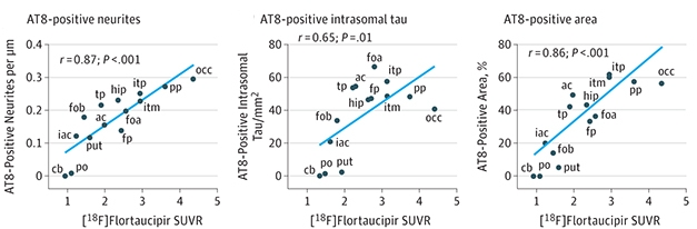
Life and Death. Uptake of flortaucipir in life matches with postmortem AT8 staining in neurites, soma, or overall in15 brain regions in a man with early onset AD. [© 2018 American Medical Association. All rights reserved.]
In this case study, the single subject was a man in his 40s with autosomal-dominant Alzheimer’s due to the PSEN1 T116N mutation. Carriers of this aggressive allele experience first symptoms in their 30s and die within four to eight years.
The man, who developed memory problems in his 30s, had two flortaucipir PET scans 20 months apart. The first, at 43, revealed tracer uptake in posterior brain regions (Smith et al., 2016). From there, his disease progressed rapidly, as did tau accumulation. On a second scan only 20 months later, tauopathy had spread to his frontal cortex and to the anterior parts of the temporal lobe, with annual increases of 20 percent to 40 percent in some parts of the brain. The man died of pneumonia one year after his last scan.
The researchers measured tau pathology postmortem either by immunostaining with the anti-phosphotau antibody AT8, or with Gallyas silver staining, in 14 discrete brain regions. They counted the number of tau-positive neurites (neuropil threads) or intrasomal neurofibrillary tangles per area, or took a measure of total tau load by tallying colored pixels on images of the stained tissue. Both antibody and silver staining revealed extensive tau-positive neurites and intrasomal tangles, with the highest density in the occipital cortex, the site of early PET uptake.
Whether they looked at neuritic tau, somal tau, or total tau burden, using AT8 or silver stain, the scientists found that tau pathology in different brain regions correlated significantly with the flortaucipir standard value uptake ratio (SUVR). The correlation with intrasomal AT8-postive tau tangles was slightly worse than the other measures, but still significant. Importantly, flortaucipir did not pick up amyloid pathology: The investigators found no correlation between the extent of amyloid plaques at autopsy and regional flortaucipir SUVR.
The results strongly support the idea that flortaucipir PET uptake is due mainly to tau pathology. Nonetheless, the work describes but one patient, whose aggressive form of AD advanced with a distinct pattern of tau spreading compared with typical late-onset AD. The authors do not think this changes the fundamental relationship between tau pathology and flortaucipir uptake, and believe their results will carry over to other forms of late onset AD.
The new study suggests that the total PET signal results from binding to both tangles and neuropil threads (Braak and Braak, 1995). However, a precise distinction between detection of neuropil threads and tangles by flortaucipir in vivo is hard to make with the techniques at hand, Lowe wrote. To nail that down, researchers should compare the PET signal in regions with predominantly neuritic or somal pathology, but that is difficult given that such areas are small and fall below the spatial resolution of PET.—Pat McCaffrey
References
News Citations
- Tau PET Aligns Spread of Pathology with Alzheimer’s Staging
- It’s Official: Tau PET Sees Tangles, and Staging Tangles Predicts Decline
Mutations Citations
Paper Citations
- Marquié M, Siao Tick Chong M, Antón-Fernández A, Verwer EE, Sáez-Calveras N, Meltzer AC, Ramanan P, Amaral AC, Gonzalez J, Normandin MD, Frosch MP, Gómez-Isla T. [F-18]-AV-1451 binding correlates with postmortem neurofibrillary tangle Braak staging. Acta Neuropathol. 2017 Oct;134(4):619-628. Epub 2017 Jun 13 PubMed.
- Smith R, Wibom M, Olsson T, Hägerström D, Jögi J, Rabinovici GD, Hansson O. Posterior Accumulation of Tau and Concordant Hypometabolism in an Early-Onset Alzheimer's Disease Patient with Presenilin-1 Mutation. J Alzheimers Dis. 2016;51(2):339-43. PubMed.
- Braak H, Braak E. Staging of Alzheimer's disease-related neurofibrillary changes. Neurobiol Aging. 1995 May-Jun;16(3):271-8; discussion 278-84. PubMed.
Further Reading
No Available Further Reading
Primary Papers
- Smith R, Wibom M, Pawlik D, Englund E, Hansson O. Correlation of In Vivo [18F]Flortaucipir With Postmortem Alzheimer Disease Tau Pathology. JAMA Neurol. 2018 Dec 3; PubMed.
Annotate
To make an annotation you must Login or Register.
Comments
Mayo Clinic
This is a brilliant study that shows us just how well Flortaucipir PET matches neuropathologically mapped tau. The correlations were strong as tested in a wide variety brain regions. The study clearly adds to our knowledge of Flortaucipir PET performance.
In my mind, the question of somal versus neuritic tau association with Flortaucipir PET is not completely answered. It does not appear that somal-predominant or neuritic-predominant tau regions were compared directly to in vivo flortaucipir PET signal for the same regions … and not that that is possible with in vivo flortaucipir PET. There could easily be cross contamination on the in vivo flortaucipir PET signal between such regions, as it would be in the mm and submillimeter range—let alone getting acceptable image registration with the autopsy tissue at this scale. This is a monumental challenge that runs up against our limitations of physics.
Despite this limitation, the correlation with flortaucipir PET overall is still a great story.
Make a Comment
To make a comment you must login or register.