Are CSF Assays Finally Ready for Prime Time?
Quick Links
This story was updated on 18 April 2017 to correct an error.
Cerebrospinal fluid biomarkers of Alzheimer’s disease have transformed clinical research, but transferring this technology to the clinic has not been easy. Even after intense efforts at quality control, biomarker readings using laboratory-grade assays swing widely between different labs, batches, and runs. Part of the problem can be solved by turning to automated systems, which tighten up biomarker readings from different runs within a few percent of each other. Even so, research labs still set their own biomarker cutoffs due to differences in the pre-analytical handling of samples. This made it impossible to agree on a single global cutoff for a biomarker level that indicates disease, as will be necessary to advance these markers into widespread diagnostic use.
The goal of a single cutoff has just gotten closer. Now, for the first time, researchers have validated a biomarker cutoff obtained in one cohort in a second, independent cohort. At the 13th International Conference on Alzheimer’s and Parkinson’s Diseases, held March 29 to April 2 in Vienna, Austria, Oskar Hansson of Lund University, Sweden, described how cutoffs initially set in the Swedish BioFINDER (Biomarkers for Identifying Neurodegenerative Disorders Early and Reliably) study were transferred to ADNI.
The Swedish group first derived a correction factor based on differences in how the respective BioFINDER and ADNI protocols handle CSF before plunking the tubes into a machine for analysis. Then the researchers used an automated assay to determine the cutoffs that predicted amyloid positivity in the BioFINDER cohort, applied the correction factor, and found that the same cutoffs correctly identified ADNI participants with brain amyloid accumulation. This represents the first clinical validation of automated CSF assays, Hansson noted. “These assays can now be considered for routine clinical use,” he told Alzforum.
The talk was enthusiastically received by both academic and pharmaceutical researchers in attendance. “It’s a very impressive study,” said Michael Weiner of the University of California, San Francisco, who oversees ADNI. John Sims of Eli Lilly in Indianapolis noted that this work will facilitate the running of worldwide trials. Right now, trials have to ship all CSF samples to the same analytical site to reduce measurement variability, greatly increasing time and expense.
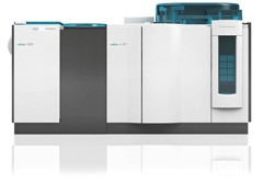
Robot Repeatability.
Use of automated systems like this one to analyze CSF samples has brought variation down to within standards of routine clinical chemistry. [Courtesy of Tobias Bittner, Roche.]
The development is the latest advance in an ongoing, years-long international effort to improve and standardize CSF measurements. Kaj Blennow of the University of Gothenburg, Sweden, leads the Global Biomarker Standardization Consortium. The consortium previously reported that despite its best efforts to standardize CSF assay protocols, identical samples run at different centers still vary by around 20 percent (see Mattsson et al., 2013). Using an automated system can bring this inter-lab variability down below 4 percent, meeting diagnostic standards (see Aug 2015 conference news). The machines do nothing to standardize the pre-analytical handling of samples, however.
To tackle this source of variation, Hansson, Blennow, and colleagues partnered with Roche Diagnostics, Basel, Switzerland. Roche makes the Elecsys immunoassays for Aβ42, total tau, and phosphotau. They run on the Cobas E601 machine that is used for routine clinical analyses across medicine, including cardiovascular care, diabetes, oncology, and infectious disease (see image above). Readings on this instrument have been shown to correlate closely with results obtained by a mass-spectrometry-based reference measurement protocol (see Bittner et al., 2016). Other companies, including Fujirebio Diagnostics, have developed similar systems.
Hansson and colleagues selected 277 BioFINDER participants with mild cognitive symptoms who had undergone both lumbar punctures and amyloid PET scans with the tracer flutemetamol. CSF samples were analyzed by Elecsys to find the cutoffs that separated amyloid-positive from amyloid-negative participants with 90 percent sensitivity. For Aβ42, this value turned out to be 1,100 pg/ml. While values lower than this reliably picked up people with brain amyloid, this cutoff also produced numerous false positives, with a diagnostic specificity of only 72 percent. By taking the ratio of either total tau or phosphotau to Aβ42 that achieved 90 percent sensitivity instead, the researchers improved the diagnostic specificity to 89 percent. The p-tau/Aβ42 cutoff was 0.022.
Notably, this 1,100 pg/ml Aβ42 cutoff is almost six times greater than the previously reported value of 192 pg/ml, obtained with the AlzBio3 assay, which distinguished people with autopsy-confirmed AD from healthy controls (see Aug 2010 news). Tobias Bittner of Roche attributes the different numbers to differences in standardization of the requisite assays. Elecsys was standardized to the LCMS-based reference method (Leinenbach et al., 2014), whereas the Alzbio3 assay was standardized based on weighted Aβ42 material. “Both cutpoints reflect the perfect clinical separation of amyloid-positive from amyloid-negative samples; just the numerical values are different,” Bittner wrote to Alzforum.
The Swedish researchers then compared CSF handling protocols between BioFINDER and ADNI. In Vienna, Hansson noted that because Aβ sticks to the sides of plastic tubes, a large amount of the protein can be lost just by aliquotting it into smaller tubes. A poster from Eline Willemse of VU University Medical Center, Amsterdam, in collaboration with the diagnostics company Euroimmun in Luebeck, Germany, corroborated this, reporting a 5-10 percent loss of Aβ every time CSF is transferred between tubes. Notably, the ADNI protocol involved many more transfer steps than BioFINDER’s. To quantify the loss, the researchers performed a small study on CSF obtained from 20 people undergoing lumbar punctures to treat normal-pressure hydrocephalus. The researchers split each sample in half and processed one half according to the BioFINDER protocol, the other ADNI’s. The ADNI protocol reliably produced peptide values about 20 percent lower than BioFINDER’s did. Thus, the researchers obtained a correction factor of 0.8 for converting Aβ42 cutoffs from BioFINDER to ADNI. Tau levels, on the other hand, did not significantly vary between the two cohorts.
Applying the correction factor, the researchers obtained cutoff values for the ADNI cohort of 880 pg/ml Aβ42, or a p-tau/Aβ42 ratio of 0.028. They used these cutoffs to stratify 646 ADNI participants who had either subjective memory complaints, mild cognitive impairment, or AD. All had undergone florbetapir amyloid PET scans. The corrected BioFINDER p-tau/Aβ42 cutoff separated ADNI amyloid-positive and amyloid-negative participants with a sensitivity of 88 percent and a specificity of 93 percent. The findings demonstrate that cutoffs from one cohort can be used diagnostically in another, Hansson said.
In both cohorts, the p-tau/Aβ42 and t-tau/Aβ42 ratios agreed with amyloid PET imaging 90 percent of the time. This is the maximum possible concordance with PET, since scans read by different radiologists agree only 90 percent of the time. The data establish automated CSF biomarker assays as equivalent to a PET visual read, Bittner told Alzforum. The Swedish study used visual reads rather than SUVRs because the former are approved for diagnostic use. However, SUVR data produce similar results, Hansson noted in Vienna.
While the use of a conversion factor allowed comparison of BioFINDER and ADNI data, researchers agree that the ultimate goal will be to have a standardized protocol for handling CSF samples. The Global Biomarker Standardization Consortium is developing such a unified sample handling protocol. Preferably, the protocol should involve as few steps as possible. Bittner suggested that, ideally, CSF would be collected in a single tube made of low-binding plastic, and then that tube would be plopped into the machine for analysis, with no transfers, freezing, centrifugation, or other manipulation of the sample. When a clinic needs to ship samples to a lab for analysis, they could be shipped fresh within 24 hours, Bittner added.
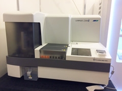
Benchtop Devices.
Companies such as Fujirebio (pictured) and Euroimmun make benchtop analyzers that run AD biomarker immunoassays. [Courtesy of Madolyn Rogers.]
This scheme would be practical because the automated systems are already in widespread use in clinics and labs worldwide. Bittner boasted that Roche has around 20,000 units in use globally, with no location more than 24 hours shipping time away from a unit.
Competing companies make similar systems. Fujirebio Diagnostics in Malvern, Pennsylvania, which acquired the Innogenetics CSF assays, runs them on a benchtop machine called Lumipulse G600II (see photo above). Hansson has worked with this system and reports similar precision to the Roche automated assays. Euroimmun also has a benchtop device, the EUROIMMUN RA Analyzer 15, that reads chemiluminescence immunoassays for Aβ42 and Aβ40.
All three companies are racing to get their AD assays to market. Roche expects to release its Aβ42 assay in June 2017, and its tau assays by the end of the year. Fujirebio reports that its assays for Aβ42 and total tau are already available in Europe, with phosphotau and Aβ40 in development. Euroimmun has finalized the technical validation for Aβ42 and Aβ40, and plans to have tau assays ready by year’s end as well. The Euroimmun assays use antibodies developed by its partner, ADx Neurosciences. All three companies will put a CE mark on their assays, indicating conformity with European standards, and are looking into requirements for approval by the U.S. Food and Drug Administration.
In addition, all three companies are validating the tests’ performance against patient samples. Manu Vandijck at Fujirebio said they have defined a normal range for Aβ42 in healthy subjects (see Wallin et al., 2015). Typical Aβ42 values in people with mild cognitive impairment or subjective cognitive decline were determined in the BioFINDER cohort, and that data has been submitted for publication, Vandijck added. Fujirebio is running studies on the other biomarkers, and also correlating CSF results with PET scanning. Britta Brix at Euroimmun noted that the company’s automated assays correlate almost perfectly (99 percent) with its manual ELISAs. Euroimmun is confirming that the biomarker cutoffs established for its ELISAs hold for the automated assays using clinical sample sets from Germany and elsewhere.
Tanja Schubert of Bioclinica Lab, a biomarker analysis company headquartered in Doylestown, Pennsylvania, performed some of the technical validation on the Euroimmun assays. To her mind, automated systems have an added advantage in that they can easily and cheaply run single patient samples. With ELISAs, each run requires a new kit, making it quite expensive to run small groups of samples, and clinical sites often wait with analysis until they have enough samples to fill a kit. The ability to run small samples quickly makes screening of participants for clinical trials more efficient, Schubert said.
One final piece to the puzzle of preparing CSF biomarkers in routine medical care will be the development of a standard CSF reference material against which all groups can calibrate their tests. That is in progress and expected to be ready later this year (see Oct 2015 news; Kuhlmann et al., 2016).—Madolyn Bowman Rogers
References
News Citations
- CSF Aβ Assays Remain Fickle: Robots to the Rescue?
- Triple Confirmation: AD Footprint in CSF of Cognitively Normal People
- Not Sexy but Oh-So-Important: Committee Blesses Way to Measure CSF Aβ
Paper Citations
- Mattsson N, Andreasson U, Persson S, Carrillo MC, Collins S, Chalbot S, Cutler N, Dufour-Rainfray D, Fagan AM, Heegaard NH, Robin Hsiung GY, Hyman B, Iqbal K, Lachno DR, Lleó A, Lewczuk P, Molinuevo JL, Parchi P, Regeniter A, Rissman R, Rosenmann H, Sancesario G, Schröder J, Shaw LM, Teunissen CE, Trojanowski JQ, Vanderstichele H, Vandijck M, Verbeek MM, Zetterberg H, Blennow K, Käser SA, Alzheimer's Association QC Program Work Group. CSF biomarker variability in the Alzheimer's Association quality control program. Alzheimers Dement. 2013 May;9(3):251-61. PubMed.
- Bittner T, Zetterberg H, Teunissen CE, Ostlund RE Jr, Militello M, Andreasson U, Hubeek I, Gibson D, Chu DC, Eichenlaub U, Heiss P, Kobold U, Leinenbach A, Madin K, Manuilova E, Rabe C, Blennow K. Technical performance of a novel, fully automated electrochemiluminescence immunoassay for the quantitation of β-amyloid (1-42) in human cerebrospinal fluid. Alzheimers Dement. 2016 May;12(5):517-26. Epub 2015 Nov 10 PubMed.
- Leinenbach A, Pannee J, Dülffer T, Huber A, Bittner T, Andreasson U, Gobom J, Zetterberg H, Kobold U, Portelius E, Blennow K, IFCC Scientific Division Working Group on CSF proteins. Mass spectrometry-based candidate reference measurement procedure for quantification of amyloid-β in cerebrospinal fluid. Clin Chem. 2014 Jul;60(7):987-94. Epub 2014 May 19 PubMed.
- Wallin A, Nordlund A, Jonsson M, Lind K, Edman Å, Göthlin M, Stålhammar J, Eckerström M, Kern S, Börjesson-Hanson A, Carlsson M, Olsson E, Zetterberg H, Blennow K, Svensson J, Öhrfelt A, Bjerke M, Rolstad S, Eckerström C. The Gothenburg MCI study: design and distribution of Alzheimer's disease and subcortical vascular disease diagnoses from baseline to 6-year follow-up. J Cereb Blood Flow Metab. 2015 Jul 15; PubMed.
- Kuhlmann J, Andreasson U, Pannee J, Bjerke M, Portelius E, Leinenbach A, Bittner T, Korecka M, Jenkins RG, Vanderstichele H, Stoops E, Lewczuk P, Shaw LM, Zegers I, Schimmel H, Zetterberg H, Blennow K, IFCC Working Group on Standardization of CSF proteins (WG-CSF). CSF Aβ1-42 - an excellent but complicated Alzheimer's biomarker - a route to standardisation. Clin Chim Acta. 2016 May 20; PubMed.
External Citations
Further Reading
Annotate
To make an annotation you must Login or Register.
Comments
No Available Comments
Make a Comment
To make a comment you must login or register.