Innovate, Then Stagnate? Brain Banks Improve Access But Are Now in Peril
Quick Links
To some, brain banks are creepy places that store and catalog frozen pieces of dead people’s central nervous systems. To neurodegenerative disease researchers, they provide a crucial service. Investigators need diseased tissue to glean insights into mechanisms and possible treatments. Pathology reports nail diagnoses, allowing clinicians to better manage new patients. Just last week, proceedings at the NIA’s Alzheimer’s Disease Research Summit 2015 were shot through with comments about the need for excellent brain banking in the field. Alas, all has not been well with brain banks of late. Just when researchers require more high-quality, well-characterized brain tissue than ever to parse the earliest stages of disease, these institutions face funding shortfalls that threaten their existence. Most brain banks store insufficient material to meet demand, and merely combining samples from several banks is not a solution because processing and storage systems are not standardized.
To overcome these shortcomings, modern banks are evolving. Globally, their curators are embracing the idea of big data and have created large databases of merged brain bank inventories. In turn, neuropathologists are grappling with the challenges of managing large data sets so information can be cataloged, stored, analyzed, and shared. They are also beginning to standardize protocols for processing tissue. These changes already are making it easier for researchers to find and match samples from different banks. Alzforum maintains a listing of consortia and individual brain banks around the world.
This series summarizes the strides that brain banks have made and the problems that remain. Part 1 covers the history, purpose, and funding of brain banks, Part 2 describes networks now in use across the world, and Part 3 delineates resources for specific diseases such as Alzheimer’s and Parkinson’s—some quite powerful and arguably underused.

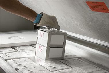
Processing Brain Donations.
Once lifted gently from a donor’s skull, half the brain is cut into thick coronal slices for flash-freezing. The other half is fixed in formalin [Images courtesy of the Wisconsin Brain Donor Program.]
Brains Undergird Big Findings
Brain banks have been around for decades, often associated with medical centers and universities. Many are small, have only a few dedicated personnel, and share morgue space, offices, and neuropathology services with hospitals. For example, the brain bank supported by the Wisconsin Alzheimer’s Disease Research Center in Madison is a typical mid-size bank with more than 300 donor samples. It employs three full-time staff (a manager, coordinator, and pathologist), and rents space in a secure research wing of the William S. Middleton Veterans Memorial Hospital. Tissue, blood, and cerebrospinal fluid samples are housed in several freezer units. Here, bright pink slabs of brain rest in labeled boxes, safely maintained at 80 degrees below zero Celsius behind frosty inner doors. The brain bank uses facilities at the new University of Wisconsin Hospital’s morgue, a brightly lit room where gleaming stainless-steel tables and weighing scales provide cold contrast to the human organs pickling in formalin in jars on the shelves.
Given their modest size, brain banks have wielded enormous impact. Not only do their autopsies confirm diagnoses and help train residents and medical students, but the study of stored tissue has produced breakthroughs in neurodegenerative disease research. “The discovery of the amyloid β fragment was first made in postmortem frozen tissue,” pointed out Nigel Cairns, a neuropathologist at Washington University in St. Louis, Missouri (Glenner and Wong, 1984). Likewise, banked tissue helped identify α-synuclein in Parkinson’s disease, tau in frontotemporal dementia (FTD), and TDP-43 in amyotrophic lateral sclerosis (ALS). More recently, researchers used genetic material from banked cases of FTD to identify the C9ORF72 gene, a major cause of ALS and FTD (Sep 2011 news).
With that, have all the important discoveries been made? Au contraire, neurologists say. As they analyze banked brains, neurologists continue to discover previously unrecognized diseases, such as senile complex tauopathy (Kovacs et al., 2011), and subtypes of known diseases, for example a variant of FTD caused by a mutation in the PRKAR1B gene (Wong et al., 2014). Importantly, understanding variants of Alzheimer’s, such as the hippocampal-sparing form, or evolving concepts, like primary age-related tauopathy, aka PART, would be unthinkable without good material from brain banks for researchers to study (Murray et al., 2011; Crary et al., 2014).
As molecular brain imaging is advancing rapidly with the discovery of PET tracers for tau, microglial activation, and amyloid beta, autopsy tissue is essential to validate what the tracers are actually binding and how to use them for differential diagnosis. If anything, brain imagers and neuropathologists are tightening their dialogue. For example, two of the three invited keynote lectures at the Human Amyloid Imaging meeting last month in Miami Beach, Florida, were by neuropathologists who are drawing heavily on brain banks to advance research on tauopathies (play lectures at HAI; Feb 2015 conference news).
Brain banks can expose problems with current diagnostic standards. For example, neuropathologist Thomas Beach at Banner Sun Health Research Institute, Sun City, Arizona, compared clinical and pathological diagnoses on banked brains from 919 dementia patients. Even though the patients had been seen at specialty clinics, their AD diagnoses were correct in only about 70 percent of cases (Beach et al., 2012). The reliability of clinical diagnoses directly affects the success of trials, said Walter Schulz-Schaeffer, a neuropathologist at the University of Göttingen, Germany. “For each 10 percent of diagnoses that are incorrect, we have to double the study group to get significant results,” he told Alzforum.
Findings facilitated by brain banks can quite directly improve patient treatment. For example, autoimmune encephalitis, a disorder in which the immune system attacks the brain, causes memory and speech problems and is often mistaken for dementia, said Annemieke Rozemuller, a neuropathologist at the Netherlands Brain Bank in Amsterdam. The disease can be treated with anti-inflammatory drugs if it’s caught early, but if misdiagnosed and untreated, it can lead to lasting brain damage. By studying banked brain tissue and associated clinical data from people with autoimmune encephalitis, Rozemuller was able to pinpoint biomarker and brain scan changes that mark the condition. She then trained neurologists how to recognize the disorder, leading to faster and more effective treatment for many patients.
Cold Storage.
Untreated brain slices are kept at 80 degrees below zero Celsius. Researchers extract proteins and DNA from these samples for biochemical and genetic experiments. [Photograph courtesy of the Netherlands Brain Bank, Netherlands Institute for Neuroscience, Amsterdam.]
Pathology studies also point toward new therapeutic strategies. Schulz-Schaeffer wanted to know how α-synuclein damages neurons in Parkinson’s disease. By studying donated brain tissue from patients, he found that more than 90 percent of α-synuclein aggregates occurred not in Lewy bodies, but in microaggregates at individual presynaptic terminals. At these synapses, dendritic spines retracted, breaking the connection between nerve cells (see Feb 2007 news story on Kramer and Schulz-Schaeffer, 2007). The finding suggests that Parkinson’s could be better treated in the long run by rescuing synapses rather than by simply replacing neurotransmitters, Schulz-Schaeffer said.
Each such finding sets off new research. The present drugs for treating Alzheimer’s were identified based on studies of postmortem human brain tissue more than 30 years ago, Beach noted in a commentary in the Journal of Alzheimer's Disease. However, since that time, funding for studies that use human brain tissue has dropped to one-tenth of what it once was, Beach writes. He even blames this waning support for the “Valley of Death,” the field’s much-deplored inability to carry basic research findings forward to new approved treatments (Beach, 2013). “It may be because of this funding decline that we are not discovering new therapeutic approaches at the rate we used to,” Beach wrote to Alzforum.
As Money Dries Up, Future Looms Uncertain
Neuropathologists around the world told Alzforum that funding represents the biggest current challenge for brain banks. European banks in particular are suffering, as grants from the European Commission have run out. Banks now are expected to support themselves by charging user fees, supplemented with local support from hospitals and universities and grants from neuroscience institutes. “We are struggling to pay for the basics now,” said Schulz-Schaeffer. Likewise, government support for Australian banks ended last year. Banks in the United States still enjoy some public funding, but it does not cover everything, and directors must tap multiple other monetary streams to make up for the shortfall. Increasingly, banks are eyeing collaborations with the deeper pockets of industry, Rozemuller said. This move brings its own caveats, as brain banks strive to keep their materials freely available to all researchers (Graeber, 2008).
The funding shortage curtails brain banks’ ability to acquire new tissue. An autopsy can run to $10,000, including the costs of transporting the body, dissecting and processing the brain, conducting complex neuropathologic assessment, and storing material, Cairns told Alzforum. Ironically, many banks are forced to turn away brain donations just as researchers need more material. “At the moment in time when science is exploding, the funding is contracting,” said Thomas Montine at the University of Washington, Seattle. “We have built these precious resources with taxpayer money over the last 30 years, but it takes time and money to maintain what we have, and even more to grow them.”
This means that researchers who request tissue have to modify their experiments to use fewer samples than they had planned, or investigate a different brain area because some, like the hippocampus, are both small and in high demand. “There is not enough tissue to meet all of the research requests. We could be doing more definitive experiments if we had greater resources,” Montine noted.
Brain banks face other difficulties as well. Most banks need more brain donations from normal controls of all ages, as well as from minorities and from people at preclinical stages of disease. Simply encouraging more donations from the public will not solve the problem, however. That’s because the most valuable samples come from people who have participated in longitudinal studies of aging or memory. Such donations are a precious capstone on extensive cognitive and clinical data that allow researchers to correlate pathology with the symptoms and biomarker changes the person had during life. Therefore brain banks prioritize these autopsies over “walk-ins,” and many willing donors have trouble finding a brain bank that will take them. People interested in donating can join a longitudinal study, but few such studies exist because they are expensive.
Last but not least, lack of money for training has led to a dearth of young neuropathologists in Europe. “We grow grayer and grayer, and nobody is coming to replace us,” said Rozemuller. In the last 50 years, the number of neuropathologists on staff at the Netherlands Brain Bank has dropped from 65 to about 15, she added.—Madolyn Rogers
References
News Citations
- Corrupt Code: DNA Repeats Are Common Cause for ALS and FTD
- Dopamine Neurons Take the Fall When Scaffolding Collapses
Conference Coverage Series Citations
Paper Citations
- Glenner GG, Wong CW. Alzheimer's disease: initial report of the purification and characterization of a novel cerebrovascular amyloid protein. Biochem Biophys Res Commun. 1984 May 16;120(3):885-90. PubMed.
- Kovacs GG, Molnár K, László L, Ströbel T, Botond G, Hönigschnabl S, Reiner-Concin A, Palkovits M, Fischer P, Budka H. A peculiar constellation of tau pathology defines a subset of dementia in the elderly. Acta Neuropathol. 2011 Aug;122(2):205-22. PubMed.
- Wong TH, Chiu WZ, Breedveld GJ, Li KW, Verkerk AJ, Hondius D, Hukema RK, Seelaar H, Frick P, Severijnen LA, Lammers GJ, Lebbink JH, van Duinen SG, Kamphorst W, Rozemuller AJ, Netherlands Brain Bank, Bakker EB, International Parkinsonism Genetics Network, Neumann M, Willemsen R, Bonifati V, Smit AB, van Swieten J. PRKAR1B mutation associated with a new neurodegenerative disorder with unique pathology. Brain. 2014 May;137(Pt 5):1361-73. Epub 2014 Apr 9 PubMed.
- Murray ME, Graff-Radford NR, Ross OA, Petersen RC, Duara R, Dickson DW. Neuropathologically defined subtypes of Alzheimer's disease with distinct clinical characteristics: a retrospective study. Lancet Neurol. 2011 Sep;10(9):785-96. PubMed.
- Crary JF, Trojanowski JQ, Schneider JA, Abisambra JF, Abner EL, Alafuzoff I, Arnold SE, Attems J, Beach TG, Bigio EH, Cairns NJ, Dickson DW, Gearing M, Grinberg LT, Hof PR, Hyman BT, Jellinger K, Jicha GA, Kovacs GG, Knopman DS, Kofler J, Kukull WA, Mackenzie IR, Masliah E, McKee A, Montine TJ, Murray ME, Neltner JH, Santa-Maria I, Seeley WW, Serrano-Pozo A, Shelanski ML, Stein T, Takao M, Thal DR, Toledo JB, Troncoso JC, Vonsattel JP, White CL 3rd, Wisniewski T, Woltjer RL, Yamada M, Nelson PT. Primary age-related tauopathy (PART): a common pathology associated with human aging. Acta Neuropathol. 2014 Dec;128(6):755-66. Epub 2014 Oct 28 PubMed.
- Beach TG, Monsell SE, Phillips LE, Kukull W. Accuracy of the clinical diagnosis of Alzheimer disease at National Institute on Aging Alzheimer Disease Centers, 2005-2010. J Neuropathol Exp Neurol. 2012 Apr;71(4):266-73. PubMed.
- Kramer ML, Schulz-Schaeffer WJ. Presynaptic alpha-synuclein aggregates, not Lewy bodies, cause neurodegeneration in dementia with Lewy bodies. J Neurosci. 2007 Feb 7;27(6):1405-10. PubMed.
- Beach TG. Alzheimer's disease and the "Valley Of Death": not enough guidance from human brain tissue?. J Alzheimers Dis. 2013;33 Suppl 1:S219-33. PubMed.
- Graeber MB. Twenty-first century brain banking: at the crossroads. Acta Neuropathol. 2008 May;115(5):493-6. PubMed.
Other Citations
External Citations
Further Reading
Annotate
To make an annotation you must Login or Register.
Comments
No Available Comments
Make a Comment
To make a comment you must login or register.