Stress and Trauma: Blast Injuries in the Military
Quick Links
Scientists are beginning to get a handle on what happens in the brains of athletes who suffer multiple concussions, but to date little research exists on how brains respond to the pressure waves from explosions. Elaine Peskind at the VA Puget Sound Health Care System, Seattle, and the University of Washington, Seattle, is gathering data on what kind of physical changes blast damage can cause in people. In a VA-funded project, she has been examining a group of 35 Iraq veterans who have each been exposed to multiple explosions. The soldiers are not cognitively impaired, but complain of memory problems, forgetfulness, and trouble concentrating. As a group, they perform about one-half to one standard deviation below age- and education-matched norms, Peskind said.
Peskind employs four neuroimaging techniques, two functional and two structural. All four techniques show brain changes in blast-exposed veterans compared to Iraq veterans without such exposure, Peskind said. Using positron emission tomography with the glucose analog fluorodeoxyglucose F18 (FDG-PET) to visualize the brain’s glucose metabolism, Peskind found lower metabolism in the posterior cingulate and parietal lobes in blast-exposed soldiers compared to Iraq-deployed controls, Peskind said. She noted that the pattern of changes is similar to that seen in early-stage mild cognitive impairment (MCI), although much less severe. Similarly, functional MRI imaging also showed MCI-like changes, with lowered activity in posterior cingulate cortex and parietal lobes. Both blast-exposed and non-blast-exposed veterans had hypometabolism in pons and cerebellum compared to civilian controls, as seen by FDG-PET (see Peskind et al., 2011).
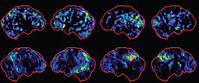
Brains with mild TBI (top row) show a similar pattern of changes to MCI brains (bottom row) by PET scan.Image credit: Elaine Peskind
The structural techniques provided more detailed data on the nature of the changes and revealed axon damage. Diffusion tensor imaging, which measures the random movement of water in the brain, exposed flaws in the integrity of white-matter tracts, particularly in the cerebellum, Peskind said. Although FDG-PET showed lower metabolism in the cerebellum in all Iraq veterans, diffusion tensor imaging revealed worse cerebellar abnormalities in the blast-exposed veterans. The second structural technique, cross-relaxation imaging, gives an estimate of the health and density of myelin, as well as of changes in gray matter (see, e.g., Yarnykh and Yuan, 2004). Peskind said this technique revealed pronounced loss of structural integrity in both white and gray matter in blast-exposed veterans compared to unexposed colleagues.
The neuroimaging data still need to be replicated, Peskind cautioned. Even so, they raise potential concerns. Is repetitive blast trauma similar to repetitive impact trauma? If so, are veterans at risk for the neurodegenerative condition of chronic traumatic encephalopathy, seen in some athletes? Will veterans with these early brain changes have an increased risk of Alzheimer’s disease later in life? “Those are my concerns, but it’s certainly too early to draw any conclusions about it,” Peskind said.
Peskind hopes to follow these veterans, whose mean age is 34, over many years and monitor how their brains change. This data will be correlated with regular neuropsychological test results. Peskind’s group is also collecting cerebrospinal fluid (CSF) from the veterans to measure alterations in the levels of biomarkers such as Aβ42. They do not yet have enough CSF data to analyze, Peskind said. She is not doing any amyloid PET scanning, but Peskind points out that Aβ levels in CSF have been shown to fall before amyloid deposits become visible in the brain. Therefore, the researchers expect to catch early amyloid changes in the CSF. The veterans have also contributed DNA for genotyping several genes of interest: apolipoprotein E, the AD risk-modifying gene TOMM40, the tau gene MAPT, and brain-derived neurotrophic factor.
In the near term, cross-relaxation imaging shows good potential as a diagnostic, Peskind said, because the analysis is simple and the finding robust. In contrast, conventional MRI or CT scans are not useful to diagnose early TBI-related brain changes, points out David Brody of Washington University in St. Louis, Missouri. “Those techniques are just not sensitive enough to detect axonal injury,” Brody said.
Peskind’s data show global changes in brain structure and function, but what is happening at the cellular level in brains exposed to blasts? The shock wave of an explosion becomes a compression wave in the brain, subjecting cells to extreme pressure. In another VA-funded project, researchers led by Pamela VandeVord at the John D. Dingell VA Medical Center, Detroit, Michigan, and Wayne State University, Detroit, are developing models to answer this question (see VA story). VandeVord’s team uses a device called a barochamber to subject brain cell cultures to high pressure. The researchers have found elevated astrocyte reactivity in the hours following a pressure wave (see VandeVord et al., 2008 and Leung et al., 2008). Astrocytes respond by highly expressing genes involved in cell survival immediately after a blast, but minimal apoptosis. In contrast, neurons ramped up apoptotic genes in the first three days after a pressure wave, indicating these fragile cells were dying (Leung and VandeVord, 2009).
The Detroit researchers also expose rats to pressure waves by forcing a blast of air down a 22-foot-long metal tube, simulating what happens during an explosion. Initial experiments showed that this technique works to increase intracranial pressure, and that older rats with thicker skulls actually get hammered hardest, suggesting that the skull helps transmit pressure (see Leonardi et al., 2011). In ongoing work, the scientists will examine the rats with MRI, cognitive testing, and blood tests for biomarkers to dissect the effects of pressure on living brains. The researchers eventually hope to use these techniques to develop therapeutics to ameliorate the effects of blasts.
Such models may help to fill a gap in the field. McKee notes that one of the primary research needs is for better models of brain injury that accurately replicate human pathology. “We do not have a perfect model yet,” she said. For example, in one study of mild repetitive impact brain injury, only one out of 18 mice developed the expected neurofibrillary tangle pathology (see Yoshiyama et al., 2005).
Better animal models should help scientists find more effective treatments. To date, animal trials have translated poorly to people. For example, in mice, a statin called simvastatin reduced Aβ burden and improved cognition after TBI (see Abrahamson et al., 2009), implying it could reduce long-term AD risk, but human treatment trials of statins in AD so far have been unsuccessful. In the future, anti-tau therapies (which aren’t anywhere near regulatory approval yet) might show promise, McKee suggested, since tau pathology is prominent in CTE brains. Henrik Zetterberg at Sahlgrenska University Hospital in Molndal, Sweden, speculates that drugs that stimulate axon outgrowth and inhibit scar formation might be beneficial.
At the moment, “There is no standard of care other than symptomatic management,” Peskind said. McKee said much of the current focus for athletes is on prevention. By taking athletes out of action for a longer period of time after a mild brain injury, physicians hope to avoid some of the long-term complications from repeated trauma. The military is adopting the same procedures for wounded soldiers and marines, McKee said.
McKee sees some treatment potential in the finding that brain changes are already apparent in young athletes exposed to repetitive concussive injuries. “Hopefully, if we can get a handle on what causes those initial changes, we can prevent this disease from spreading into full-blown disease,” she said. All researchers interviewed for this story agreed that much more research is needed. Peskind summed it up: “We are in the infancy of studying a truly new and unique phenomenon.” For an overview of PTSD and dementia, check out Part 5.—Madolyn Bowman Rogers.
This is Part 4 of a six-part series. See also Part 1, Part 2, Part 3, Part 5, Part 6.
References
News Citations
- Stress and Trauma: The Puzzle of Post-traumatic Stress Disorder
- Stress and Trauma: New Frontier Lures Alzheimer’s Researchers
- Stress and Trauma: Aβ’s Mysterious Role in Severe Brain Injury
- Stress and Trauma: Shaken Brains, Shaken Lives
- Stress and Trauma: Tackling Post-traumatic Stress Disorder
Paper Citations
- Peskind ER, Petrie EC, Cross DJ, Pagulayan K, McCraw K, Hoff D, Hart K, Yu CE, Raskind MA, Cook DG, Minoshima S. Cerebrocerebellar hypometabolism associated with repetitive blast exposure mild traumatic brain injury in 12 Iraq war Veterans with persistent post-concussive symptoms. Neuroimage. 2011 Jan;54 Suppl 1:S76-82. PubMed.
- Yarnykh VL, Yuan C. Cross-relaxation imaging reveals detailed anatomy of white matter fiber tracts in the human brain. Neuroimage. 2004 Sep;23(1):409-24. PubMed.
- Vandevord PJ, Leung LY, Hardy W, Mason M, Yang KH, King AI. Up-regulation of reactivity and survival genes in astrocytes after exposure to short duration overpressure. Neurosci Lett. 2008 Apr 4;434(3):247-52. PubMed.
- Leung LY, Vandevord PJ, Dal Cengio AL, Bir C, Yang KH, King AI. Blast related neurotrauma: a review of cellular injury. Mol Cell Biomech. 2008 Sep;5(3):155-68. PubMed.
- Leonardi AD, Bir CA, Ritzel DV, Vandevord PJ. Intracranial pressure increases during exposure to a shock wave. J Neurotrauma. 2011 Jan;28(1):85-94. PubMed.
- Yoshiyama Y, Uryu K, Higuchi M, Longhi L, Hoover R, Fujimoto S, McIntosh T, Lee VM, Trojanowski JQ. Enhanced neurofibrillary tangle formation, cerebral atrophy, and cognitive deficits induced by repetitive mild brain injury in a transgenic tauopathy mouse model. J Neurotrauma. 2005 Oct;22(10):1134-41. PubMed.
- Abrahamson EE, Ikonomovic MD, Dixon CE, Dekosky ST. Simvastatin therapy prevents brain trauma-induced increases in beta-amyloid peptide levels. Ann Neurol. 2009 Sep;66(3):407-14. PubMed.
External Citations
Further Reading
Annotate
To make an annotation you must Login or Register.
Comments
�
Such interesting reports. These researchers may have not only revealed a truly new and unique phenomenon, but also given rise to some new ideas on the etiological research of various dementias.
Looking forward to reading the other parts of this report.
Make a Comment
To make a comment you must login or register.