Research Brief: New Hues for Calcium Imagers
Quick Links
Calcium imaging aficionados, are you sick of viewing fields of green ad nauseam? Relief may be at hand, according to a paper that describes new red, blue, and improved green Ca2+ indicators for visualizing intracellular signaling. Reporting today in Science online, researchers led by Robert Campbell at the University of Alberta, Edmonton, Canada, used a bacterial screening approach to boost the sensitivity of existing calcium sensors and make them fluoresce in different colors. The new reagents improve single-color calcium imaging and enable simultaneous visualization of different compartments within single cells, opening the door to Ca2+ imaging experiments “previously impractical,” write the authors.
Campbell and first author Yongxin Zhao collaborated with scientists in Japan on a strategy to screen for improved versions of genetically encoded calcium sensors (GCaMP). First developed in 2001 (Nakai et al., 2001), GCaMPs consist of a modified green fluorescent protein (GFP) fused to peptides that bind calmodulin in the presence of calcium, making the normally dim GFP light up. In the current paper, the researchers used error-prone PCR to create mutants of GCaMP3 (Tian et al., 2009), which they expressed in E. coli bacteria and grew on plates. Screening some 200,000 colonies, they identified three variants that lit up in response to calcium about twice as brightly as GCaMP3. By varying external calcium concentration and other conditions, the researchers were able to shift the bacteria toward Ca2+-free and Ca2+-bound states, making it easier to detect Ca2+-dependent changes.
Seeing that the screen worked, the researchers tweaked the GFP constructs further in an attempt to make them light up at red or blue frequencies, instead of green. They tested the new indicators in rat hippocampal neurons (see image of red probe below) and transgenic C. elegans worms, and showed they could use their red calcium sensor in conjunction with an ATP indicator in HeLa cells. “This palette of indicators paints the way to a colorful new era of Ca2+ imaging,” the authors write.—Esther Landhuis.
Click on this image to launch the video.
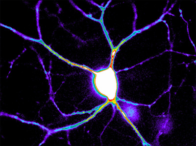
Rat hippocampal neuron expresses new red fluorescent calcium indicator. Calcium levels, from highest to lowest, appear in white, red, yellow, green, blue, and black. Image credit: Mado Lemieux and Paul De Koninck, Laval University, Quebec, CanadaVideo shows time-lapse 3-color fluorescence imaging of HeLa cells transfected with plasmids encoding new calcium sensors. Calcium can be simultaneously visualized in the nucleus (red), cytoplasm (green), and mitochondria (blue-green). Video credit: Takeharu Nagai
References
Paper Citations
- Nakai J, Ohkura M, Imoto K. A high signal-to-noise Ca(2+) probe composed of a single green fluorescent protein. Nat Biotechnol. 2001 Feb;19(2):137-41. PubMed.
- Tian L, Hires SA, Mao T, Huber D, Chiappe ME, Chalasani SH, Petreanu L, Akerboom J, McKinney SA, Schreiter ER, Bargmann CI, Jayaraman V, Svoboda K, Looger LL. Imaging neural activity in worms, flies and mice with improved GCaMP calcium indicators. Nat Methods. 2009 Dec;6(12):875-81. PubMed.
Other Citations
External Citations
Further Reading
Papers
- Nakai J, Ohkura M, Imoto K. A high signal-to-noise Ca(2+) probe composed of a single green fluorescent protein. Nat Biotechnol. 2001 Feb;19(2):137-41. PubMed.
- Tian L, Hires SA, Mao T, Huber D, Chiappe ME, Chalasani SH, Petreanu L, Akerboom J, McKinney SA, Schreiter ER, Bargmann CI, Jayaraman V, Svoboda K, Looger LL. Imaging neural activity in worms, flies and mice with improved GCaMP calcium indicators. Nat Methods. 2009 Dec;6(12):875-81. PubMed.
Primary Papers
- Zhao Y, Araki S, Wu J, Teramoto T, Chang YF, Nakano M, Abdelfattah AS, Fujiwara M, Ishihara T, Nagai T, Campbell RE. An expanded palette of genetically encoded Ca²⁺ indicators. Science. 2011 Sep 30;333(6051):1888-91. PubMed.
Annotate
To make an annotation you must Login or Register.
Comments
No Available Comments
Make a Comment
To make a comment you must login or register.