The Skinny on FAT: APP’s Role in Fast Axonal Transport
Quick Links
In neurons, APP-laden vesicles powered by kinesin motors ride from the cell body out to synapses. But is APP just a passenger, or does the protein sit in the driver’s seat? New work from Elaine Bearer of Brown University in Providence, Rhode Island, and the Marine Biological Laboratory at Woods Hole, Massachusetts, suggests the latter. In a study presented this month at the 36th annual meeting of the Society for Neuroscience (SfN) in Atlanta, Georgia, and published shortly thereafter in the October 24 PNAS online, Bearer and lead authors Prasanna Satpute-Krishnan, Joseph DeGiorgis and Michel Conley show that a 15 amino acid C-terminal peptide from human APP can recruit cargoes of synthetic beads to the fast axonal transport (FAT) machinery in squid giant axons. The results suggest that one physiological function of APP could be to direct axonal transport of vesicles or other cargoes, a process known to be compromised in Alzheimer disease (for a recent review see Stokin and Goldstein, 2006).
The squid giant axon was also on display at another presentation at the SfN meeting, this one by Gustavo Pigino from Scott Brady’s lab at the University of Illinois in Chicago. Following up on recent work showing that polyglutamate-expanded proteins inhibit FAT in extruded squid axoplasm (see ARF related news story), Pigino showed that Aβ peptide oligomers inhibit both anterograde and retrograde transport in the same system, an effect they attributed to Aβ’s activation of casein kinase II.
APP first came to the notice of Bearer and colleagues in their studies on the transport of herpes simplex virus (HSV) in neurons. The virus moves from cell body to synapse via kinesin-powered FAT. When the Brown researchers injected purified human HSV particles into isolated squid giant axons, they could reconstitute this transport, and found that the transported virus contained high amounts of human APP (see ARF related news story). APP has been implicated as a motor receptor in vesicle movement by the work of Lawrence Goldstein and others (see Kamal et al., 2000 and ARF related news story), leading Bearer to hypothesize a similar role for APP in virus transport.
This is where the study gets colorful. Rather than follow the movement of virus particles, which contain other proteins and lipids in addition to APP, the researchers decided to simplify the system. They replaced the virus with similarly sized fluorescent polystyrene beads to which they coupled defined peptides. This allowed them to test sequence requirements for transport. In this system, video microscopy of injected beads derivatized with a peptide corresponding to the last 15 amino acids of the C-terminal cytoplasmic tail of APP revealed a rapid, unidirectional anterograde transport. The speed of movement was consistent with FAT, and comparable to that seen for endogenous organelles or intact virus in the same system. The transport function was specific to the C-terminal peptide, because in the same axons, different color beads coupled to a peptide derived from the extracellular domain of APP, or a scrambled C-terminal sequence, displayed no motility at all (see movie below).
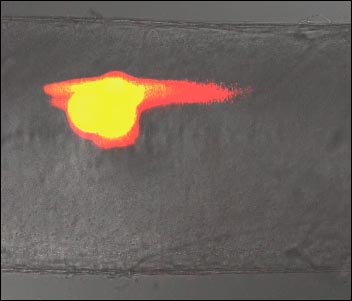
APP C-terminal Drives Fast Axonal Transport
Fluorescent virus-sized particles coupled to the C-terminal (red) of APP drive fast anterograde transport when injected into the squid giant axon, while particles coupled to the N-terminal of APP (green) remain stationary. The movie and the paper are available as open access on the PNAS website. [Image courtesy PNAS]
The function of APP in transport appears to be highly conserved. Cloning of squid APP revealed a nearly identical C-terminal sequence (13 out of 15 amino acid match to the human), and transport studies showed that the squid cytoplasmic domain also supported bead transport. In further support for a physiological role of APP in transport, the researchers demonstrated that the intact C99 fragment of human APP, consisting of a small luminal/extracellular stretch, plus the transmembrane and cytoplasmic domain, could mediate transport when conjugated to beads at a similar density as found on HSV. Transport of beads coupled to either C99 or the C-terminal peptide was blocked by addition of excess free peptide, suggesting that the protein was binding specifically to some part of the motor assembly.
The results support the idea that APP is involved in HSV transport in neurons, and raises the possibility that it could play a wider role in recruitment of the transport machinery for anterograde shipments in general. The domain contains an NPXY motif which participates in many protein-protein interactions, and could contact kinesin directly, as Goldstein has proposed, or via a scaffolding protein (Taru et al., 2002). Bearer and colleagues call the short peptide a “zipcode” for anterograde transport, but while the sequence is sufficient for transport, further work will be required to show if it is necessary.
Gustavo Pigino also showed data on FAT in the squid axon, where he and his colleagues are studying the effects of Aβ oligomers. Like Bearer, Brady and colleagues summer in Woods Hole, where they study axonal transport not in intact axons, but in extruded axoplasm preparations. Because they have removed the plasma membrane barrier, their system allows them to add proteins or inhibitors directly to cytosolic transport machinery, and look for effects on anterograde or retrograde transport and the motor proteins involved. Earlier this year, they showed that polyglutamine-expanded androgen receptor inhibits FAT by turning on jun N-terminal kinase, a regulator of kinesin activity (see ARF related news story).
In his talk, Pigino described similar experiments testing the effects of Aβ—soluble, oligomeric, or fibrillar. Only oligomeric Aβ inhibited transport at very low concentrations (100 nM), and it slowed movement in both anterograde and retrograde directions. Addition of active casein kinase II (CKII) mimicked the effect of Aβ, suggesting that the oligomers were causing activation of this enzyme. Consistent with these results, Aβ oligomers increased phosphorylation of the CKII substrate kinesin light chain, while addition of a CKII inhibitor along with Aβ restored transport. One attendee raised the issue of whether Aβ oligomers are present in the cytoplasm of neurons in sufficient amounts to elicit these effects in intact cells, a question which remains to be answered for both normal and AD brain.—Pat McCaffrey
References
News Citations
- JNK Clogs Axonal Transport
- Herpes and AD—Virus Hitches Ride with APP
- Suspects for Aβ Generation Spotted Together, En Route to Nerve Terminal
Paper Citations
- Stokin GB, Goldstein LS. Axonal Transport and Alzheimer's Disease. Annu Rev Biochem. 2006 Mar 16; PubMed.
- Kamal A, Stokin GB, Yang Z, Xia CH, Goldstein LS. Axonal transport of amyloid precursor protein is mediated by direct binding to the kinesin light chain subunit of kinesin-I. Neuron. 2000 Nov;28(2):449-59. PubMed.
- Taru H, Iijima K, Hase M, Kirino Y, Yagi Y, Suzuki T. Interaction of Alzheimer's beta -amyloid precursor family proteins with scaffold proteins of the JNK signaling cascade. J Biol Chem. 2002 May 31;277(22):20070-8. PubMed.
External Citations
Further Reading
Primary Papers
- Satpute-Krishnan P, DeGiorgis JA, Conley MP, Jang M, Bearer EL. A peptide zipcode sufficient for anterograde transport within amyloid precursor protein. Proc Natl Acad Sci U S A. 2006 Oct 31;103(44):16532-7. PubMed.
Annotate
To make an annotation you must Login or Register.
Comments
University of Pennsylvania
This is an important study that may usher in a new perspective on the normal function of APP and clarify its role in AD pathogenesis.
View all comments by John TrojanowskiUniversity of Texas Health Science Center at San Antonio
The study of the biology of APP and its proteolytic products, although pioneered in the early 1990s by Eddie Koo, Joseph Buxbaum, Sam Sisodia, and others, has nevertheless remained mostly out of the limelight until the last few years. The present study from Elaine Bearer’s laboratory now illuminates part of a picture that has been taking shape in the last few years suggesting that APP is likely involved in the modulation of synaptic activity in adults (Priller et al., 2006; Yang et al., 2005; Seabrook et al., 1999), in synapse formation and function (Wang et al., 2005), and in neuronal migration and adhesion during development (Herms et al., 2004).
APP is a synaptic protein that is anterogradely transported to terminals. A few years ago Kamal et al. suggested that the C-terminus of APP could serve as a receptor for kinesin (Kamal et al., 2000), but this observation was subsequently questioned by Lazarov et al. (Lazarov et al., 2005). The present study by Satpute-Krishnan et al. provides strong evidence that the C-terminus of APP may indeed contain sequences sufficient for its association with axonal transport components. The careful experiments addressed this question using a fairly well-defined system, the squid giant axon, and the investigators’ observations indicate that the C-terminal domain of APP, either through a direct interaction with kinesin or indirectly via scaffolding proteins such as JIPs, participates in fast anterograde axonal transport. Quoting their discussion, “The robust motility of C99 beads in the intact axon argues for a physiological role of APP in recruitment of anterograde transport machinery inside cells.” It certainly does, and it comes as no surprise. Although the study by Satpute-Krishnan et al. does not answer the question of whether the interaction of APP with kinesin is or is not direct, it significantly adds to the rapidly growing evidence suggesting a crucial role of the C-terminus of APP (and possibly its family members APLP1 and 2) in neuronal biology, possibly at synaptic sites.
The remarkable conservation of the C-terminal sequences of APP across phyla suggests conservation of function. Supporting this idea, the phenotypes of APP/APLP2 double and APP/APLP1/APLP2 triple knockouts and those of two prominent APP-interacting proteins (X11 and the Fe65 family) involve alterations in neuronal function, synaptic formation, function, and regulation (Wang et al., 2005; Ho et al., 2003; Yang et al., 2005; Priller et al., 2006), and in the case of the Fe65/FE65L double and APP/APLP1/APLP2 triple knockouts, result in cortical dysplasias and heterotopias (Herms et al., 2004, Guenette et al., 2006). Interestingly, it was recently shown that transgenic expression of AICD in combination with Fe65 causes alterations in signaling (Ryan and Pimplikar, 2005) and activation of proteins involved in growth cone collapse and axonal guidance.
Why is this important? Most of all, because a significant component of amyloid-β toxicity requires multimerization of APP and cleavage of its C-terminus at Asp664 (Lu et al., 2003; Lu et al., 2003; Shaked et al., 2006). This cleavage not only releases a toxic peptide, but also removes the sequences required for the formation of a multiplicity of protein complexes at APP’s cytoplasmic domain, and as Satpute-Krishnan now suggest, for fast axonal transport. Consistent with what may be an important role of the extreme C-terminal sequences of APP in transducing amyloid-β toxicity, we recently showed that stabilization of APP’s cytoplasmic tail by mutation of the Asp664 cleavage site had a dramatic effect in the development of AD-like deficits in transgenic mice (Galvan et al., 2006)—even in the presence of abundant amyloid-β. With this in mind, the question arises as to whether cleavage at Asp664 while in transit towards synaptic sites would, as expected, prevent delivery of the molecule to its destination—and if the hypothesis of Kamal et al. is correct, whether it would affect the delivery of any subset of associated axonal transport vesicles. Thus, a population of Asp664-intact (transport-competent) and Asp664-cleaved (transport-incompetent) APP molecules may exist. Satpute-Krishnan et al. estimate that 3,000 copies of APP may be associated with each motile bead in their system; although in this study they don’t address the question of what is the minimal number of APP molecules required for transport, it is conceivable that transport-incompetent (Asp664-cleaved) APP molecules may be “carried along” in vesicles containing a sufficient number of transport-competent (Asp664-intact) APP. Cleavage of APP at Asp664 would thus affect not only the transport-competence (and thus the rate of delivery) of APP to neuronal terminals, but since the motifs required for the interaction of APP with a variety of cellular functions reside downstream of Asp664, it would also affect the overall signaling ability of populations of APP molecules at their destination at synaptic sites.
References:
Priller C, Bauer T, Mitteregger G, Krebs B, Kretzschmar HA, Herms J. Synapse formation and function is modulated by the amyloid precursor protein. J Neurosci. 2006 Jul 5;26(27):7212-21. PubMed.
Yang G, Gong YD, Gong K, Jiang WL, Kwon E, Wang P, Zheng H, Zhang XF, Gan WB, Zhao NM. Reduced synaptic vesicle density and active zone size in mice lacking amyloid precursor protein (APP) and APP-like protein 2. Neurosci Lett. 2005 Aug 12-19;384(1-2):66-71. PubMed.
Seabrook GR, Smith DW, Bowery BJ, Easter A, Reynolds T, Fitzjohn SM, Morton RA, Zheng H, Dawson GR, Sirinathsinghji DJ, Davies CH, Collingridge GL, Hill RG. Mechanisms contributing to the deficits in hippocampal synaptic plasticity in mice lacking amyloid precursor protein. Neuropharmacology. 1999 Mar;38(3):349-59. PubMed.
Wang P, Yang G, Mosier DR, Chang P, Zaidi T, Gong YD, Zhao NM, Dominguez B, Lee KF, Gan WB, Zheng H. Defective neuromuscular synapses in mice lacking amyloid precursor protein (APP) and APP-Like protein 2. J Neurosci. 2005 Feb 2;25(5):1219-25. PubMed.
Herms J, Anliker B, Heber S, Ring S, Fuhrmann M, Kretzschmar H, Sisodia S, Müller U. Cortical dysplasia resembling human type 2 lissencephaly in mice lacking all three APP family members. EMBO J. 2004 Oct 13;23(20):4106-15. PubMed.
Kamal A, Stokin GB, Yang Z, Xia CH, Goldstein LS. Axonal transport of amyloid precursor protein is mediated by direct binding to the kinesin light chain subunit of kinesin-I. Neuron. 2000 Nov;28(2):449-59. PubMed.
Lazarov O, Morfini GA, Lee EB, Farah MH, Szodorai A, DeBoer SR, Koliatsos VE, Kins S, Lee VM, Wong PC, Price DL, Brady ST, Sisodia SS. Axonal transport, amyloid precursor protein, kinesin-1, and the processing apparatus: revisited. J Neurosci. 2005 Mar 2;25(9):2386-95. PubMed.
Ho A, Morishita W, Hammer RE, Malenka RC, Sudhof TC. A role for Mints in transmitter release: Mint 1 knockout mice exhibit impaired GABAergic synaptic transmission. Proc Natl Acad Sci U S A. 2003 Feb 4;100(3):1409-14. Epub 2003 Jan 23 PubMed.
Guénette S, Chang Y, Hiesberger T, Richardson JA, Eckman CB, Eckman EA, Hammer RE, Herz J. Essential roles for the FE65 amyloid precursor protein-interacting proteins in brain development. EMBO J. 2006 Jan 25;25(2):420-31. PubMed.
Ryan KA, Pimplikar SW. Activation of GSK-3 and phosphorylation of CRMP2 in transgenic mice expressing APP intracellular domain. J Cell Biol. 2005 Oct 24;171(2):327-35. PubMed.
Lu DC, Soriano S, Bredesen DE, Koo EH. Caspase cleavage of the amyloid precursor protein modulates amyloid beta-protein toxicity. J Neurochem. 2003 Nov;87(3):733-41. PubMed.
Lu DC, Shaked GM, Masliah E, Bredesen DE, Koo EH. Amyloid beta protein toxicity mediated by the formation of amyloid-beta protein precursor complexes. Ann Neurol. 2003 Dec;54(6):781-9. PubMed.
Shaked GM, Kummer MP, Lu DC, Galvan V, Bredesen DE, Koo EH. Abeta induces cell death by direct interaction with its cognate extracellular domain on APP (APP 597-624). FASEB J. 2006 Jun;20(8):1254-6. PubMed.
Galvan V, Gorostiza OF, Banwait S, Ataie M, Logvinova AV, Sitaraman S, Carlson E, Sagi SA, Chevallier N, Jin K, Greenberg DA, Bredesen DE. Reversal of Alzheimer's-like pathology and behavior in human APP transgenic mice by mutation of Asp664. Proc Natl Acad Sci U S A. 2006 May 2;103(18):7130-5. PubMed.
Make a Comment
To make a comment you must login or register.