New Genetics Frontiers: Finding Modifiers, Making Sense of Pathways
Quick Links
The laundry list of potential Alzheimer’s risk genes that have turned up in genome-wide association studies explains but a fraction of the total genetic burden of the disease, so geneticists are turning to whole-genome and exome sequencing to hunt down more suspects. At the 12th International Conference on Alzheimer’s and Parkinson’s Diseases, held March 18-22 in Nice, France, speakers reported on the latest progress in this area, including a potential modifier gene identified in a large Colombian kindred with early onset disease. In parallel, geneticists are moving beyond listing genes. They are now analyzing genetic interactions to determine the underlying metabolic pathways most affected by each disorder. This provides clues as to why particular neuronal populations succumb in different neurodegenerative conditions, said John Hardy of University College London. Based on data from a number of such diseases, Hardy proposed specific metabolic processes that break down in different neuronal subtypes. “I hope that understanding selective vulnerability will be the first step to being able to compensate for it,” he told the audience.
For Alzheimer’s disease, the 30 or so loci identified to date account for about 30 percent of the genetic risk, Julie Williams of Cardiff University, U.K., said in her talk. GWAS have found mostly common genes that add little risk, so much of the missing heritability may lie in rare genes with larger effects. To find them, researchers are mining the genomes of families with inherited disease. Even when the causal mutation is already known, this approach can reveal separate factors that bring on disease earlier or later, said Kenneth Kosik of the University of California, Santa Barbara. In the Colombian kindred who carry the E280A PS1 mutation, most carriers develop mild cognitive impairment as defined by MCI criteria by the age of 44, and full-blown dementia five years later (Acosta-Baena et al., 2011). However, a few carriers have long intrigued researchers because they break the pattern, succumbing either much earlier or much later.
Kosik wanted to find out which genes are behind this. He analyzed whole-genome sequences from 117 members of this kindred. That huge dataset will likely reveal more insights, but even a first analysis turned up a protective haplotype of 56 kb that delayed disease onset by about 10 years. The haplotype was common in the kindred, occurring in about one in four people. It spanned a region of cytokine genes, and contained 22 SNPs that associated with delayed onset. Only one of these fell within the coding region of a gene, however. This SNP, rs1129844, marked eotaxin-1, also called CCL11.

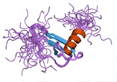
Eotaxin. Good guy? Bad guy? Genetics raises the question.
Eotaxin has already made a name for itself in aging research. The concentration of this cytokine rises with age in both mice and people. It is one of the potentially deleterious aging factors identified in parabiosis research, in which young mice receive old blood (see Nov 2009 conference news; Aug 2011 news). In those studies, eotaxin suppressed the birth of new neurons and impaired learning.
The SNP Kosik found changes an alanine to a threonine at position 23, precisely where a signal peptide is cleaved from eotaxin, he noted. This site may also play a role in allowing the cytokine to bind its receptor, CCR3. Kosik wondered if the SNP might represent the functional mutation that confers protection. Cells transfected with the variant pumped out more eotaxin, suggesting a functional effect, though its direction surprised the researchers. “We might have expected a decrease in eotaxin secretion from a protective allele,” Kosik said.
Would this SNP protect other populations as well? To try to confirm the findings, Kosik collaborated with Aimee Kao at the University of California, San Francisco, to analyze 152 AD patients there. In this group also, the protective haplotype associated with later disease onset. Moreover, in people with this haplotype, eotaxin levels did not rise with age. Kosik looked for other modifiers in this group, and found an SNP in the IL4 receptor that was enriched in people who developed disease late, and absent in those who succumbed early. This SNP delayed disease by about seven years. It occurred only in people of Latino heritage. Intriguingly, ligands for IL4R can release eotaxin and induce Aβ clearance, Kosik said.
“We put forth the hypothesis that eotaxin release may mediate Aβ clearance epistatically with IL4R,” Kosik told the audience. Epistasis refers to an interaction between different genes, such that one modifies the expression or effect of the other. Low to moderate levels of eotaxin may be beneficial and higher levels deleterious, Koski suggested, adding “Having these SNPs may shift the curve toward the beneficial side.” However, he cautioned that these data are preliminary, as the populations tested were small and not powered to reach genome-wide significance. The finding needs to be confirmed in larger populations. So far, these SNPs have not shown up in GWAS results.
Other researchers are also analyzing genetic kindreds. Christine Van Broeckhoven of the VIB, University of Antwerp, Belgium, noted that even for familial AD, the causal mutation remains unknown in 90 to 95 percent of cases. She is leading genetic studies to try to pin down these genes. In a Belgium cohort of 343 familial AD cases, she found 52 separate mutations. Of these, 41 were in presenilin 1 or 2. Carriers had widely varying ages of onset, suggesting the presence of genetic modifiers, and she is looking for those now. Surprisingly, among the other 11 genes, loci linked to neurodegenerative diseases turned up frequently, in particular granulin, a risk factor for frontotemporal dementia (FTD). Some families carried more than one gene that segregated with disease; for example, one had both a PS1 mutation and a C9ORF72 expansion; the latter is linked to FTD and amyotrophic lateral sclerosis. How these additional mutations influence pathology in AD patients is unclear, Van Broeckhoven said. Neurogeneticists don’t yet know exactly what distinguishes a modifier that increases risk from a bona fide disease gene, or how common the presence of multiple disease-associated variants in a given person will turn out to be.
A complementary growing trend in genetics is that the same gene crops up in multiple diseases, for example TREM2 in both FTD and AD, and glucocerebrosidase (GBA) in both Gaucher’s and Parkinson’s, said Rita Guerreiro of UCL. This is called pleiotrophy, and exome sequencing is uncovering more such cases. By comparing affected and unaffected individuals within a family, exome sequencing can quickly home in on mutations. In 22 parent-child trios, Guerreiro’s group has found 26 confirmed de novo mutations. In one case, exome sequencing revealed that heterozygous mutations in polynucleotide kinase 3’-phosphatase caused familial ataxia in a Portuguese kindred (see Bras et al., 2015). Homozygous mutations in this gene, on the other hand, cause developmental delays, early seizures, and small brains. PNKP plays a role in repairing DNA.
As geneticists sort through associations, metabolic pathways have begun to emerge for each disease. Hardy pointed out that nearly every Alzheimer’s gene reported to date falls into one of four pathways: cholesterol metabolism, innate immunity, endosomal vesicle recycling, and protein ubiquitination. “I’d be suspicious of any new gene that didn’t fall into one of these pathways,” he said. In Parkinson’s disease, most genes are involved in lysosomal function, mitophagy, or immunity. “These pathways must mark those cellular systems that are susceptible to disease,” he suggested.
To explore this idea, Hardy looked at gene expression data for GWAS hits to find genes that are co-expressed. This analysis allowed him to define “modules” of co-regulated genes. In the case of ataxias, many GWAS hits mapped to one of two co-expression modules: calcium homeostasis or the ubiquitin proteasome system. Both of these co-expression modules occur only in the cerebellum, with the former active in Purkinje cells and the latter in granule cells (see Bettencourt et al., 2014). The motor symptoms of ataxia arise from perturbed cerebellar function. The data imply that the cells’ vulnerability arises from these particular systems. “We see two ways to get ataxia: to have disrupted calcium homeostasis or a disrupted ubiquitin proteasome,” Hardy said. Not only does each disease variant attack a different neuron type, it also has distinct clinical features, he added.
Using the same methodology, Hardy found similar patterns for other diseases, although the data were noisier. For Parkinson’s, many genes involved in mitochondrial complex I were co-expressed in dopamine neurons of the substantia nigra. They include Parkin, Pink1, and SNCA, as well as RAB39B, which was independently identified as a PD gene by another group (see Wilson et al., 2014). The finding suggests that mitochondrial function is crucial for dopaminergic neurons, and that stresses to this system precipitate PD. For dementias, including AD, FTD, dementia with Lewy bodies, and PD dementia, lysosomal gene co-expression modules turn up over and over, primarily affecting cortical pyramidal neurons, Hardy said. Meanwhile, many FTD and ALS genes, including C9ORF72, map to the ubiquitin proteasome module. These disorders target motor and cortical neurons, which may particularly rely on this waste disposal system.
“The overall suggestion of the work we’ve been doing is that each neuronal type, for reasons having to do with its function, is close to a catastrophic cliff,” Hardy said. Dysfunction in one of the genes involved in the particular task that puts the particular neuron type near that point can push the neuron over toward disease. “Selective vulnerability has to do with what catastrophic cliff the neuron is close to,” he concluded. Many genes in these modules are involved in cleaning up cellular damage, which may explain why the diseases only manifest with age, when accumulated wear and tear puts more pressure on neurons. In answer to an audience query, Hardy reiterated the idea that in many cases there may be a “second hit” that actually precipitates disease.—Madolyn Bowman Rogers
References
Mutations Citations
News Citations
- Chicago: The Vampire Principle—Young Blood Rejuvenates Aging Brain?
- Paper Alert: Do Blood-Borne Factors Control Brain Aging?
Alzpedia Citations
Paper Citations
- Acosta-Baena N, Sepulveda-Falla D, Lopera-Gómez CM, Jaramillo-Elorza MC, Moreno S, Aguirre-Acevedo DC, Saldarriaga A, Lopera F. Pre-dementia clinical stages in presenilin 1 E280A familial early-onset Alzheimer's disease: a retrospective cohort study. Lancet Neurol. 2011 Mar;10(3):213-20. PubMed.
- Bras J, Alonso I, Barbot C, Costa MM, Darwent L, Orme T, Sequeiros J, Hardy J, Coutinho P, Guerreiro R. Mutations in PNKP Cause Recessive Ataxia with Oculomotor Apraxia Type 4. Am J Hum Genet. 2015 Mar 5;96(3):474-9. Epub 2015 Feb 26 PubMed.
- Bettencourt C, Ryten M, Forabosco P, Schorge S, Hersheson J, Hardy J, Houlden H, United Kingdom Brain Expression Consortium. Insights from cerebellar transcriptomic analysis into the pathogenesis of ataxia. JAMA Neurol. 2014 Jul 1;71(7):831-9. PubMed.
- Wilson GR, Sim JC, McLean C, Giannandrea M, Galea CA, Riseley JR, Stephenson SE, Fitzpatrick E, Haas SA, Pope K, Hogan KJ, Gregg RG, Bromhead CJ, Wargowski DS, Lawrence CH, James PA, Churchyard A, Gao Y, Phelan DG, Gillies G, Salce N, Stanford L, Marsh AP, Mignogna ML, Hayflick SJ, Leventer RJ, Delatycki MB, Mellick GD, Kalscheuer VM, D'Adamo P, Bahlo M, Amor DJ, Lockhart PJ. Mutations in RAB39B cause X-linked intellectual disability and early-onset Parkinson disease with α-synuclein pathology. Am J Hum Genet. 2014 Dec 4;95(6):729-35. Epub 2014 Nov 26 PubMed.
External Citations
Further Reading
News
- 44 Parent-Child Trios Yield 40 Candidate ALS Genes
- Genentech Strikes Deal with 23andMe to Study Parkinson’s Genomes
- Researchers Build on GWAS to Parse Genetic Players in AD and PD
- Exome–Network Combination Uncovers New Disease Genes
- Late-Onset Disease Clusters in Families with Alzheimer’s
- Alzheimer’s Whole-Genome Data Now Available From the NIH
- Pooled GWAS Reveals New Alzheimer’s Genes and Pathways
- The Power of Three: Genetic Trios Yield ALS Gene Candidates
- Stream of Genetics Pushes FTD Research Forward
- Genetics Project Update: Over 1,000 Genomes and Counting
- Barcelona: What Lies Beyond Genomewide Association Studies?
Annotate
To make an annotation you must Login or Register.

Comments
No Available Comments
Make a Comment
To make a comment you must login or register.